

How Pulmonary Contusion Radiology Identifies a Bruised Lung
Pulmonary contusion radiology looks for lung bruising caused by chest trauma. On CT, the main signs are patchy ground-glass opacity or poorly defined consolidation from bleeding and swelling in the lung. These areas are usually non-segmental, often near the chest wall, and may cross normal lung fissures. A chest X-ray can miss an early contusion, so CT is more reliable when injuries are serious or symptoms are worsening.
After a car accident, imaging also checks for related injuries, including rib fractures, pneumothorax, hemothorax, and pulmonary laceration. This matters because a contusion can worsen during the first 24 hours, even when the first X-ray looks normal.
Most uncomplicated contusions begin to improve on imaging within 48 hours and clear within 10 to 14 days. Opacity that keeps worsening or fails to clear may point to aspiration, pneumonia, atelectasis, ARDS, or an underlying lung laceration.
I’m Dr. Adam Savage, and this guide will help you understand what trauma imaging can show after a chest injury and why the timing of each scan matters.
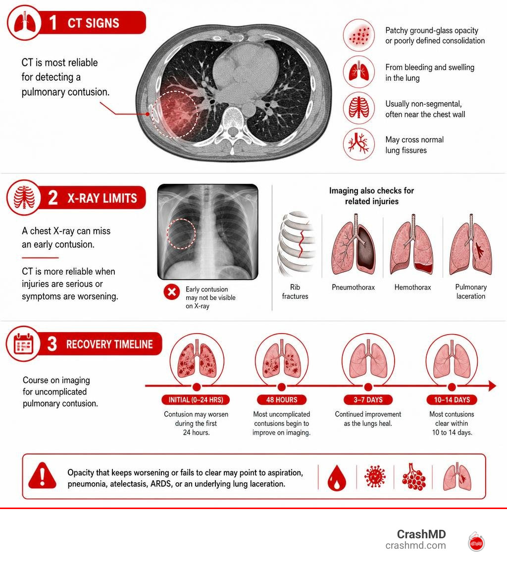
Simple pulmonary contusion radiology glossary:
Pathophysiology and Essential Hallmarks of Lung Parenchymal Trauma
A pulmonary contusion is an injury to the lung parenchyma involving microvascular disruption, alveolar capillary rupture, and interstitial edema without frank parenchymal laceration. It is the single most common parenchymal lung injury encountered in non-penetrating blunt chest trauma, seen in roughly 17% to 70% of thoracic impact cases.
When blunt kinetic energy impacts the chest wall, shockwaves compress the underlying spongy lung tissue. This pressure wave causes microvascular shear stress. As alveolar walls stretch and tear at a microscopic level, blood and serous fluid extravasate into the interstitial spaces and alveolar cavities.
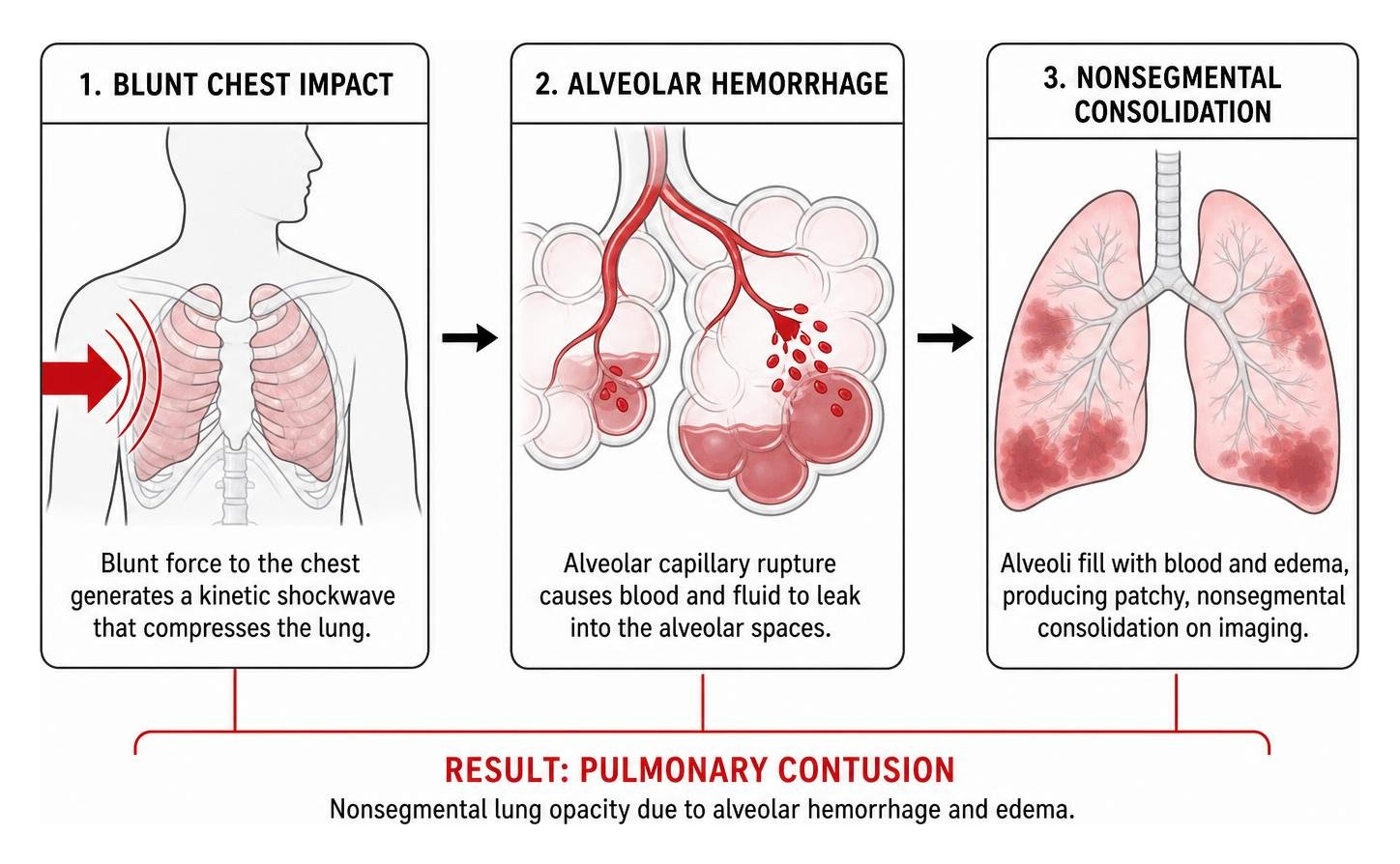
Unlike lobar pneumonia or vascular infarctions, a pulmonary contusion does not respect classical bronchoarterial segments. Its distribution is characteristically non-segmental, reflecting the physical vector of blunt force rather than vascular or bronchial anatomy. For an in-depth clinical look at mechanical tissue injury, see our step-by-step guide to internal chest bruising and explore the Radiopaedia reference guide on pulmonary contusion.
Core Modalities in Pulmonary Contusion Radiology: X-Ray vs. CT
In modern emergency departments, evaluating acute thoracic trauma involves two primary diagnostic tools: portable anteroposterior (AP) chest radiography and multidetector computed tomography (MDCT).
While a portable chest X-ray remains the rapid first-line exam in the trauma bay, MDCT serves as the gold standard for grading injury severity via systems like the American Association for the Surgery of Trauma (AAST) scale.
| Diagnostic Metric | Portable Plain Radiography (CXR) | Multidetector Computed Tomography (MDCT) |
|---|---|---|
| Early Sensitivity (<6 hours) | ~38% (frequently appears normal) | Near 100% |
| Morphological Detail | Non-specific hazy airspace density | Differentiates GGO from dense consolidation |
| Associated Lesion Detection | Misses occult pneumothorax/lacerations | Accurately identifies lacerations, effusion, occult air |
| Subpleural Assessment | Cannot resolve subpleural margins | Clearly visualizes 1–2 mm subpleural sparing |
| Clinical Utility | Rapid initial triage and tube placement | Definitive injury staging and procedural planning |
Understanding these differences helps patients understand why doctors order advanced scans even after an initial normal X-ray, as outlined in our practical guide to pulmonary contusion.
Plain Film Limitations in Pulmonary Contusion Radiology
Standard plain film radiography suffers from significant diagnostic delay. Alveolar hemorrhage and secondary inflammatory edema evolve gradually over time. Consequently, a baseline chest X-ray taken within the initial 4 to 6 hours after injury has a sensitivity as low as 38%.
On plain film, contusions present as patchy, ill-defined, non-segmental opacities. However, early imaging frequently underestimates parenchymal damage. Peak radiographic opacification typically occurs 24 hours post-trauma.
Furthermore, supine AP chest X-rays frequently obscure occult pneumothoraces and subtle rib fractures. Clinicians relying entirely on immediate plain radiographs risk underestimating the patient’s respiratory risk, a limitation detailed in the MSD Manual clinical reference for pulmonary contusion.
Advanced Multidetector CT Findings in Pulmonary Contusion Radiology
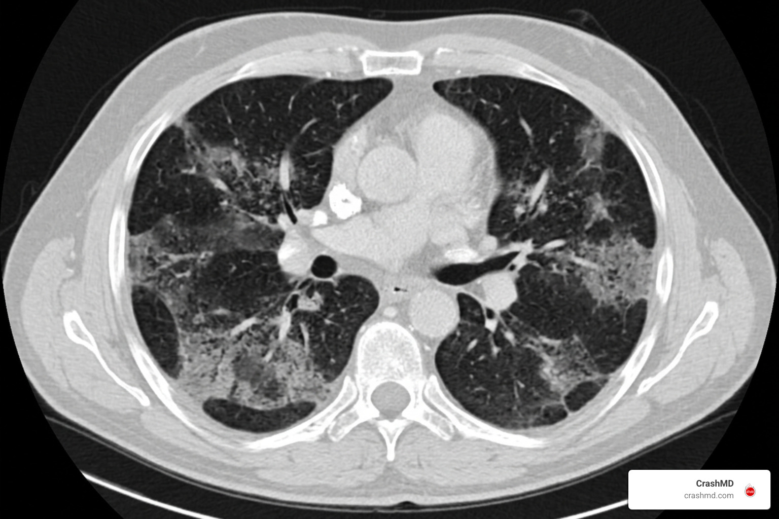
Multidetector CT provides near 100% sensitivity for blunt parenchymal damage. In a study of 93 patients evaluated with MDCT:
- 47.3% presented exclusively with ground-glass opacities (GGO), reflecting partial alveolar fluid filling and interstitial capillary engorgement.
- 52.7% showed mixed opacities or dense, homogeneous consolidation indicating extensive alveolar flooding.
- 80.6% demonstrated multiple contusion opacities rather than single isolated foci.
- 52.7% demonstrated bilateral pulmonary contusions.
CT findings typically predominate in the posterior and peripheral lung segments across all vertical lung thirds. Because distal terminal bronchioles fill with blood and inflammatory exudates, classic air bronchograms are relatively rare within severe contusions compared to infectious consolidations. Thin-slice reconstruction allows radiologists to map the exact three-dimensional volume of injured lung, as described in the Radiology Key review of chest trauma.
Distinguishing Subpleural Sparing, Fissure Crossing, and Pediatric Manifestations
Modern volumetric CT captures distinct geometric patterns caused by blunt impact dynamics. Diagnostic accuracy improves when clinicians properly map these lesions, an important consideration when evaluating billing records such as the ultimate guide to right pulmonary contusion icd-10 code.
Subpleural Sparing and Fissure Crossing Diagnostic Signs
Two distinctive radiological signs often appear on trauma CT scans:
- Subpleural Sparing: A preserved, unaffected 1 to 2 mm strip of lung parenchyma immediately beneath the visceral pleura. While historically considered ubiquitous, MDCT research demonstrates subpleural sparing is present in 26.9% of patients.
- Fissure Crossing: Non-segmental parenchymal opacification that directly crosses major or minor anatomical fissures into an adjacent pulmonary lobe without architectural interruption. This sign occurs in roughly 22.6% of cases.
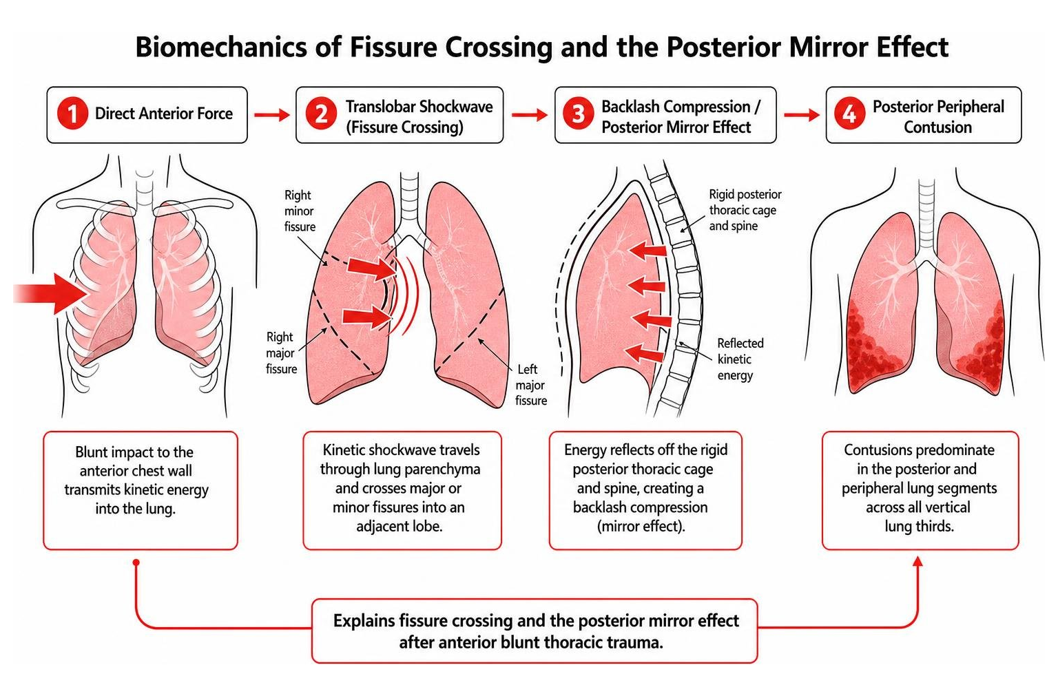
Both signs occur because kinetic shockwaves travel across lung tissue independent of pleural boundaries. Posterior lesions often develop even after frontal impacts due to the “mirror effect,” where kinetic energy reflects off the rigid posterior thoracic cage and spine. For details, see the Comparative study on multidetector CT findings.
Pediatric vs. Adult Thoracic Compliance and Injury Patterns
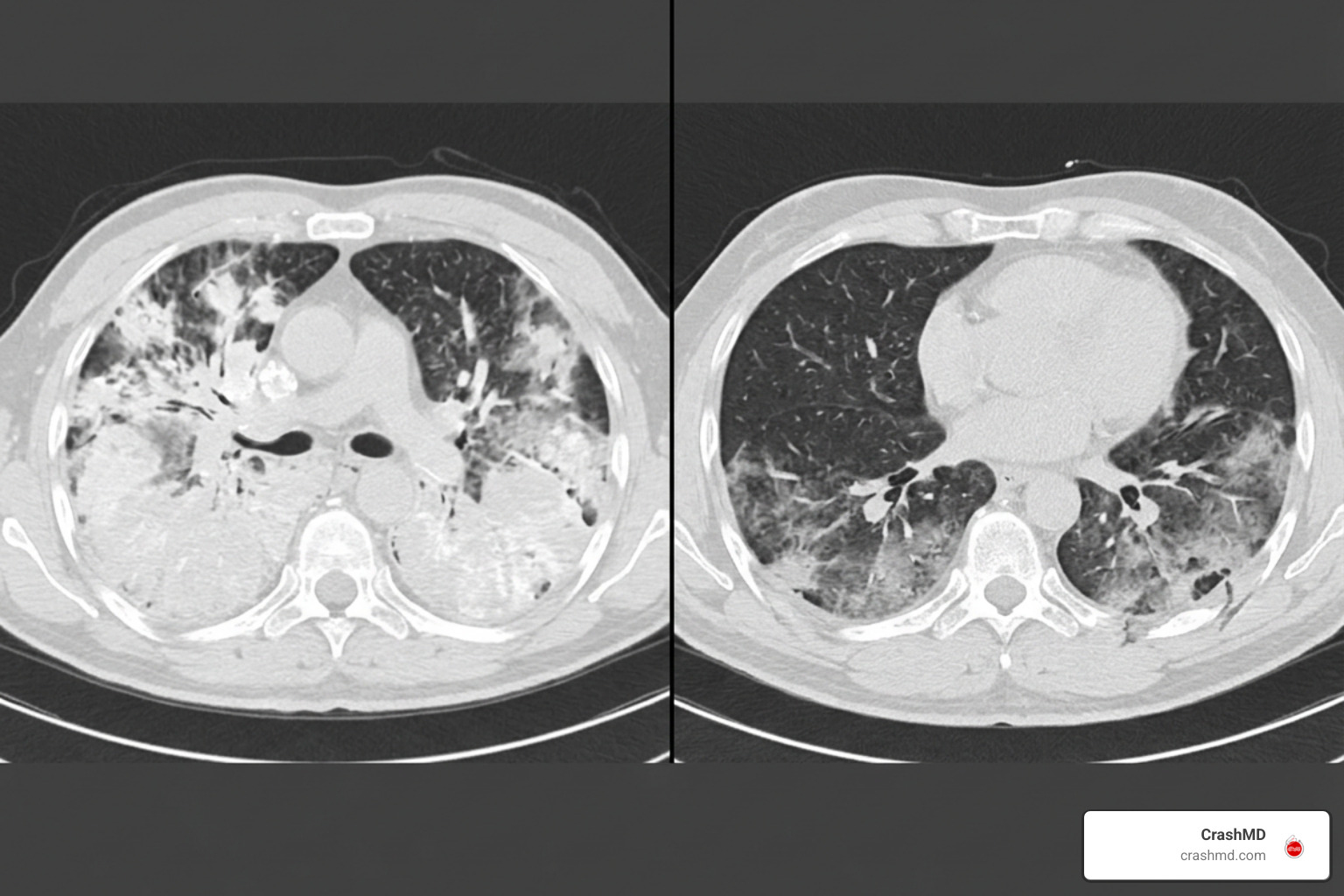
A patient’s age fundamentally alters their radiographic presentation:
- Pediatric Patients: Children possess highly compliant, uncalcified, elastic cartilaginous rib cages. Kinetic force compresses the chest wall inward and transfers directly into the pulmonary parenchyma without fracturing bones. Consequently, pediatric patients show significantly higher rates of dense, homogeneous consolidation (present in over 90% of pediatric contusions) and diffuse anteroposterior opacification.
- Adult Patients: Adults possess rigid, calcified thoracic cages that absorb energy by fracturing. Adults present predominantly with isolated ground-glass opacities (53.1% in adults vs. 8.3% in children).
A child with respiratory distress after trauma can have extensive parenchymal lung consolidation despite having zero broken ribs on their X-ray or CT scan.
Expected Healing Timeline, ARDS Risks, and Differential Diagnosis
Understanding the typical resolution timeline of a bruised lung prevents misdiagnosis and improper treatment, as highlighted in an essential guide to pulmonary contusion risks.
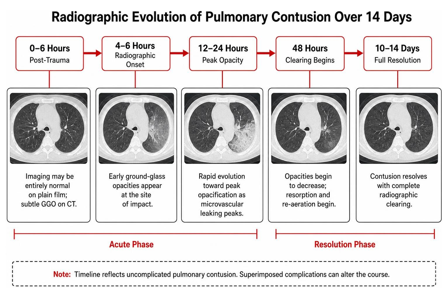
Radiographic Resolution Timeline and Superimposed Complications
An uncomplicated pulmonary contusion follows a predictable timeline:
- 0 to 6 Hours: Imaging may be entirely normal on plain film; subtle GGO on CT.
- 12 to 24 Hours: Rapid evolution toward peak opacification as microvascular leaking peaks.
- 48 Hours: Radiographic improvement and clearing should be visible.
- 10 to 14 Days: Complete radiographic resolution of parenchymal opacities.
If opacities fail to improve by day 3 to 7, or worsen after initial clearing, clinicians must evaluate for secondary complications:
- Acute Respiratory Distress Syndrome (ARDS): Diffuse, bilateral, dependent consolidations.
- Ventilator-Associated Pneumonia (VAP): High-risk complication in intubated trauma patients carrying mortality rates up to 50%.
- Unmasked Pulmonary Laceration: Resolving contusions often unmask underlying 2 to 5 cm traumatic hematomas or post-traumatic pneumatoceles (“Swiss cheese” sign) that were obscured by acute alveolar hemorrhage.
Differentiating Contusions from Aspiration, Pneumonia, and Laceration
Acute lung opacification in trauma patients requires systematic differential diagnosis:
- Bacterial Pneumonia: Characterized by progressive consolidation, prominent air bronchograms, fever, purulent sputum, and delayed onset past 48 hours.
- Aspiration Pneumonitis: Typically occupies gravitationally dependent lung regions (superior segments of lower lobes or posterior segments of upper lobes) and appears within hours of altered consciousness.
- Compressive Atelectasis: Demonstrates distinct anatomical volume loss, shifted fissures, and triangular margins.
- Fat Embolism Syndrome: Manifests 24 to 72 hours after long-bone fractures with diffuse bilateral “snowstorm” infiltrates.
Recent medical breakthroughs use artificial intelligence and radiomic texture analysis to distinguish trauma hemorrhage from bacterial exudates with over 90% accuracy, bypassing clinical diagnostic ambiguities as detailed in the AI and radiomics study in pulmonary contusion differentiation.
Associated Thoracic Injuries and Clinical Impact on Management
Because pulmonary contusions result from significant energy transfer, they rarely occur in complete isolation. Proper patient advocacy requires a thorough post-accident medical workup, as emphasized when navigating your post-accident doctor visit communication essentials.
Rib Fractures, Flail Chest, and Traumatic Pneumothorax
In adult blunt trauma cohorts, pulmonary contusions are frequently accompanied by adjacent chest wall and pleural disruptions:
- Rib Fractures (44.1%): Displaced fractures risk directly tearing visceral pleura, while undisplaced fractures are often invisible on initial plain film. Flail chest occurs when paradoxical motion of multiple fractured ribs compromises ventilation mechanics.
- Pneumothorax (44.1%): On supine trauma radiographs, look for basilar signs such as the deep sulcus sign, double diaphragm sign, or the floating fat pad sign.
- Hemothorax & Laceration (43.1% to 44.1%): High-attenuation pleural fluid (35–70 HU) exhibiting the “hematocrit sign” reflects acute bleeding, requiring multi-phase CT to rule out active intercostal artery extravasation.
Concomitant Abdominal and Vascular Injury Considerations
High-velocity blunt impacts frequently transmit energy into surrounding organ systems. Up to 83.9% of blunt trauma patients with pulmonary contusions have associated extrathoracic injuries.
Thoracic imaging protocols must evaluate for:
- Pneumomediastinum & The Macklin Effect: Alveolar rupture dissecting air centripetally along bronchovascular sheaths into the mediastinum.
- Diaphragmatic Rupture & Visceral Herniation: Commonly occurring in 8% of severe chest-abdominal trauma (predominantly left-sided).
- Solid Organ Laceration: Coexisting abdominal trauma, such as liver damage discussed in our in-depth guide to liver laceration severity and treatment.
- Thoracic Aortic Injury: Aortic pseudoaneurysms near the ductus ligamentum distal to the left subclavian artery carry high mortality and require urgent contrast-enhanced MDCT.
Frequently Asked Questions About Pulmonary Contusion Radiology
How quickly does a pulmonary contusion appear and resolve on imaging?
Signs of a pulmonary contusion appear on CT within 4 to 6 hours and peak in radiographic density at 24 hours. Normal clearing begins within 48 hours, with complete resolution expected within 10 to 14 days. Any infiltrate worsening beyond 48 hours raises suspicion for secondary infection or ARDS.
Why can children have severe pulmonary contusions without broken ribs?
Children have a flexible, cartilaginous, and uncalcified thoracic wall. This elastic compliance allows blunt kinetic force to transfer directly through the chest wall into the lung parenchyma without fracturing ribs, frequently causing extensive homogeneous consolidation.
What is the most accurate imaging test for diagnosing lung contusions?
Multidetector computed tomography (MDCT) with thin-slice reconstruction is the most accurate imaging modality. While plain chest radiographs detect only ~38% of contusions within the first 6 hours, MDCT demonstrates near 100% sensitivity for identifying subtle ground-glass opacities, subpleural sparing, and occult traumatic injuries.
Conclusion
Understanding pulmonary contusion radiology is critical for recognizing the full extent of blunt chest trauma. Because standard X-rays can miss early injury, high-resolution MDCT and serial radiographic tracking ensure that subtle alveolar hemorrhage, evolving consolidations, and underlying lacerations are accurately identified before complications arise.
At CrashMD, we guide accident victims through recovery with personalized pain management and evidence-based rehabilitation protocols across our specialized clinical practices.
If you or a loved one are recovering from a motor vehicle collision, read our comprehensive resource on car accident pulmonary contusion symptoms and recovery to ensure your health, recovery, and medical documentation are thoroughly supported.