

What a Laceration Liver Means After Trauma
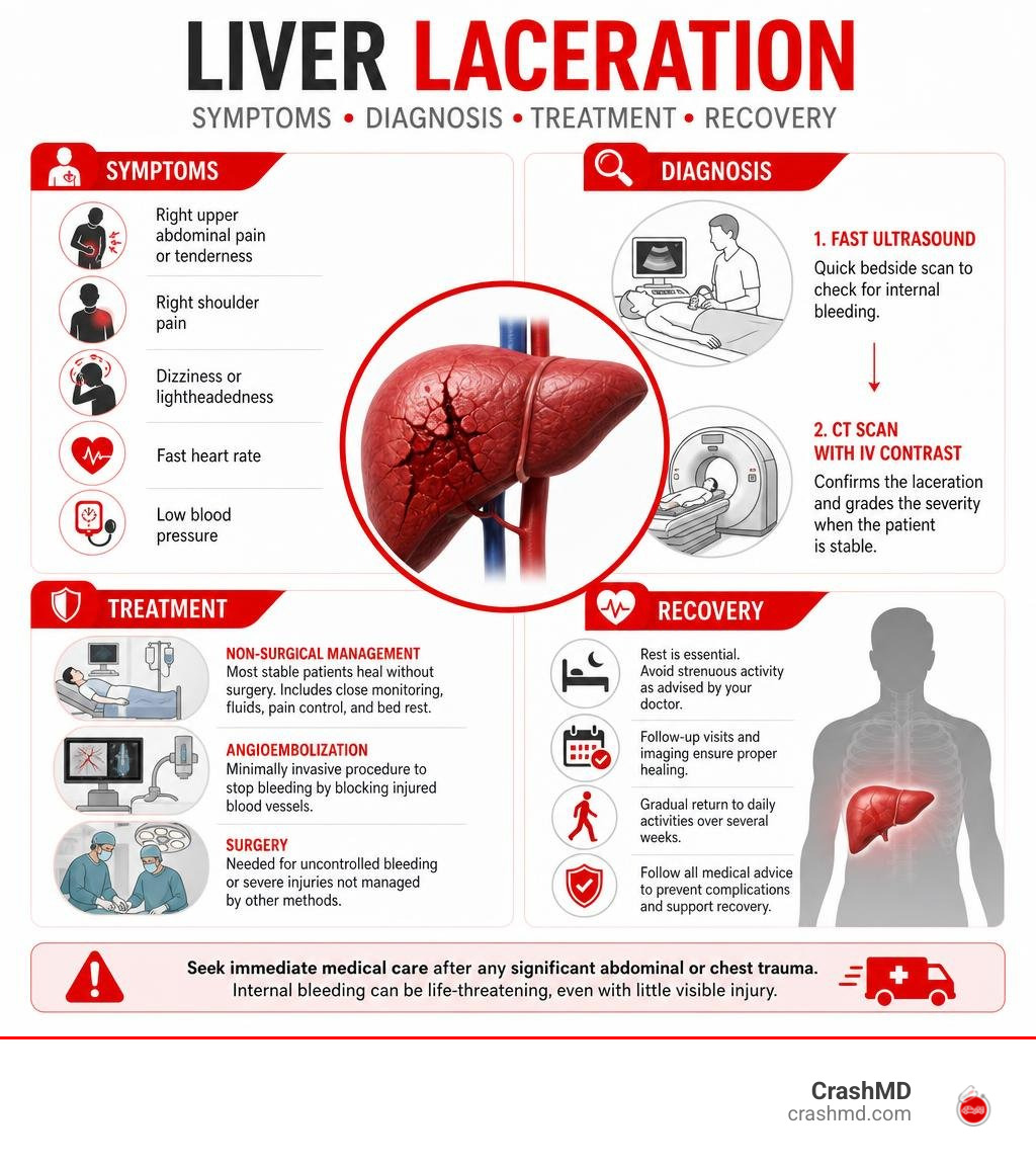
A laceration liver is a tear in the liver caused by trauma, most often a car crash, hard fall, stab wound, or gunshot wound. It can range from a small tear that heals with close hospital monitoring to a severe injury with dangerous internal bleeding that needs urgent treatment.
The liver sits high on the right side of the abdomen, beneath the ribs and diaphragm. Its large size, blood-rich tissue, and exposed position make it especially vulnerable during a sudden impact. It is the most commonly injured solid organ in blunt abdominal trauma.
| Quick answer | What it means |
|---|---|
| Common cause | Motor vehicle collisions are a leading cause of blunt liver injury. |
| Main danger | Internal bleeding can cause shock, even when there is little visible injury outside the body. |
| Common symptoms | Right upper abdominal pain, tenderness, right shoulder pain, dizziness, fast heart rate, or low blood pressure. |
| How doctors check it | Trauma teams commonly use a FAST ultrasound first, followed by a CT scan with IV contrast when the patient is stable. |
| Can it heal without surgery? | Often, yes. More than 80% of liver injuries can be managed without an operation when bleeding is controlled and vital signs remain stable. |
Liver injuries account for about 5% of trauma admissions, and most are minor. But no one should assume pain after a crash is “just bruising.” A liver tear can bleed heavily, and symptoms may worsen over time. Fast evaluation matters, especially after a high-impact collision or a direct blow to the right upper abdomen or lower chest.
I’m Dr. Adam Savage, and the sections ahead explain how liver lacerations are graded, diagnosed, treated, and followed during recovery.
Anatomical Susceptibility and Mechanisms Causing a Laceration Liver
The liver is the largest internal solid organ in the human body, anchoring the upper right quadrant of the abdomen directly beneath the diaphragm. Wrapped in a thin fibrous envelope known as Glisson’s capsule, the organ is soft, densely vascularized, and heavy. The massive right lobe occupies most of the space under the lower ribs, making it the most frequently damaged region during abdominal impact.
Because the liver shifts position during breathing—rising as high as the T4 nipple line during full expiration and descending to T12 during deep inspiration—lower thoracic chest impacts frequently transfer energy directly into hepatic tissue. Learn more about evaluating chest Wall impacts in our step-by-step guide to internal chest bruising. Interestingly, a patient can suffer a severe laceration liver injury without a single broken rib. Research into Blunt Liver Injury with Intact Ribs under Impacts on the Abdomen: A Biomechanical Investigation shows that the rib cage can flex under sudden blunt pressure, transferring concentrated physical strain to the hyperelastic liver tissue beneath before the bones themselves reach their breaking point.
High-Impact Traumatic Causes and Biomechanics
Motor vehicle collisions represent the primary cause of blunt hepatic trauma. When a vehicle halts abruptly during a crash, rapid momentum transfer causes internal organs to slam against the rigid musculoskeletal frame. Lateral or broadside impacts carry a particularly high risk because the side ribs possess greater mobility and push directly into the right hepatic lobe.
Other major injury mechanisms include:
- High-Altitude Falls: Industrial accidents or falls from roofs compress the liver between the anterior abdominal wall and the vertebral spine.
- Direct Blunt Blows: Motorcycle collisions, pedestrian impacts, or heavy machinery accidents deliver concentrated kinetic force.
- Penetrating Trauma: Gunshot wounds and stab wounds directly slice Glisson’s capsule and intrahepatic blood vessels. While penetrating gunshot wounds require surgery in roughly 90% of cases, stab wounds can sometimes be managed conservatively if vital signs remain stable.
Diagnostic Signs and Hemorrhagic Shock Symptoms
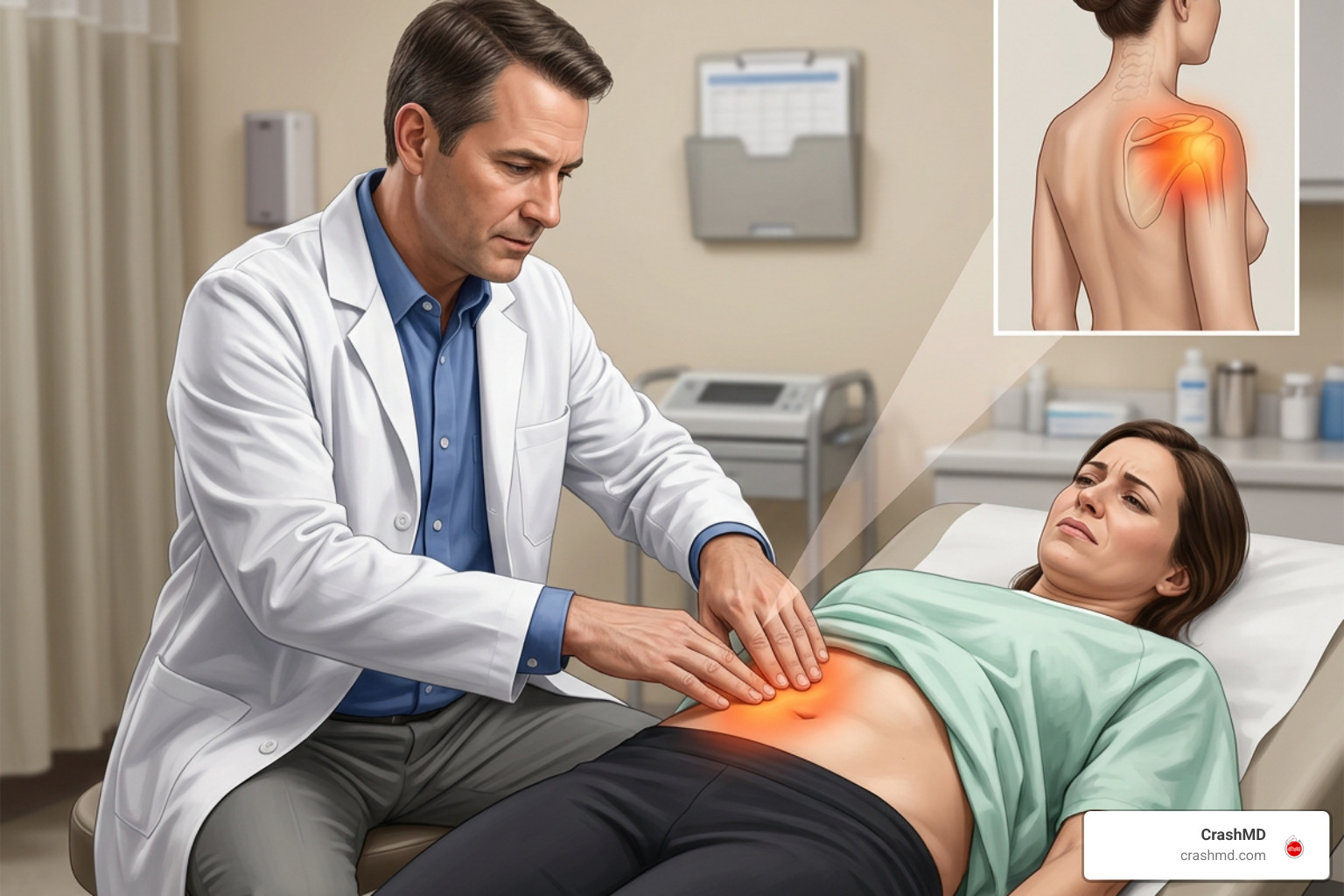
A liver laceration triggers immediate bleeding into the peritoneal cavity (hemoperitoneum). Common physical symptoms include:
- Severe, localized pain and tenderness in the right upper quadrant.
- Involuntary abdominal guarding or rigid wall stiffness.
- Kehr’s Sign: Referred pain felt at the tip of the right shoulder. Blood irritating the underside of the diaphragm stimulates the phrenic nerve, sending misdirected pain signals up to the shoulder.
If arterial or venous bleeding continues unchecked, the patient can rapidly slip into hemorrhagic shock. Signs of circulatory failure include low blood pressure (hypotension under 90 mmHg), rapid heart rate (tachycardia over 120 beats per minute), pale clammy skin, confusion, and dizziness.
Diagnostic Evaluation and Injury Grading Systems
When a trauma patient arrives at an emergency department, clinicians must quickly evaluate whether internal organs are bleeding. Initial laboratory testing routinely checks blood counts and liver function. Elevated liver enzymes—specifically ALT and AST—are 100% specific and approximately 93% sensitive for predicting liver tissue injury after blunt trauma.
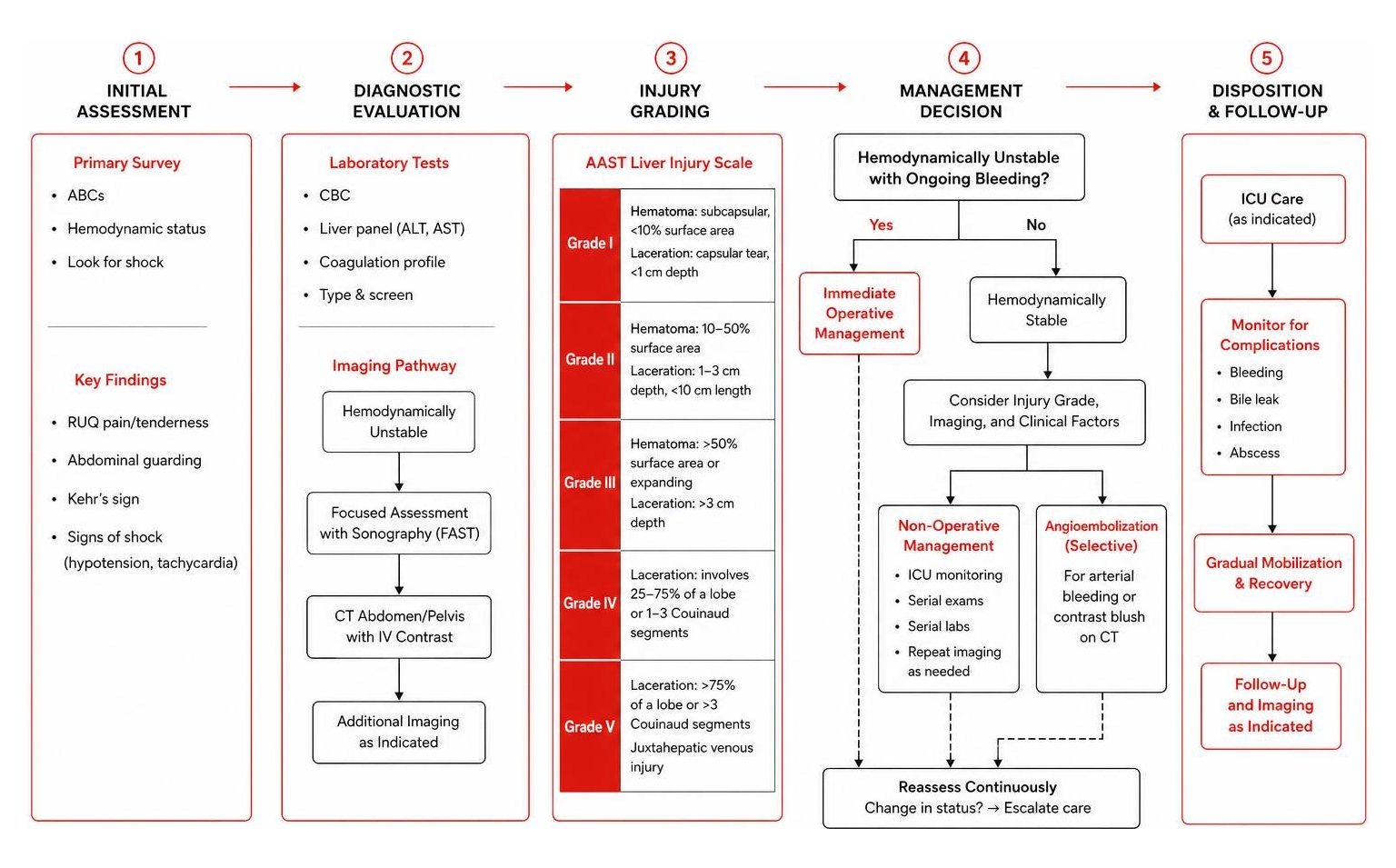
To establish an accurate anatomical diagnosis, clinicians rely on targeted imaging guidelines detailed in medical literature on the Management of liver trauma in adults – PMC – NIH.
Diagnostic Tools for Assessing a Laceration Liver
| Diagnostic Tool | Primary Purpose | Key Advantages | Clinical Limitations |
|---|---|---|---|
| FAST Ultrasound | Bedside screening for free intra-abdominal fluid. | Rapid (under 5 minutes), portable, zero radiation, safe in unstable patients. | Cannot grade laceration depth; low sensitivity in pediatric cases or small bleeds. |
| Contrast CT Scan | Gold standard imaging for stable patients. | 96%–100% accurate; identifies active arterial “blush,” hematoma volume, and anatomical grade. | Requires patient stability and transportation to radiology suite. |
| Serial Hematocrits | Laboratory monitoring of ongoing blood loss. | Tracks trends in blood volume stability over time. | Lagging indicator; does not show real-time arterial spurts. |
AAST Grading Scale for Laceration Liver Severity
The American Association for the Surgery of Trauma (AAST) classifies hepatic injuries on a scale from Grade I (least severe) to Grade VI (fatal hepatic avulsion). Anatomical disruption is often quantified using Couinaud liver segments (the eight functional anatomical subdivisions of the liver).
- Grade I: Subcapsular hematoma taking up less than 10% of surface area, or a capsular tear under 1 cm in parenchymal depth.
- Grade II: Subcapsular hematoma occupying 10% to 50% of surface area, intraparenchymal hematoma under 10 cm, or laceration 1 cm to 3 cm deep and under 10 cm in length.
- Grade III: Subcapsular hematoma greater than 50% of surface area, expanding or ruptured hematoma, intraparenchymal hematoma over 10 cm, or laceration deeper than 3 cm.
- Grade IV: Parenchymal disruption involving 25% to 75% of a hepatic lobe or 1 to 3 Couinaud segments within a single lobe.
- Grade V: Parenchymal disruption exceeding 75% of a liver lobe or major juxtahepatic venous injuries (retrohepatic vena cava or main portal vein branches).
- Grade VI: Complete hepatic avulsion, severing the liver from its primary blood supply.
Grades I and II make up 80% to 90% of hepatic injuries and carry an excellent prognosis when monitored properly.
Non-Operative vs. Operative Management Strategies
Modern trauma centers prioritize non-operative management (NOM) whenever possible. In fact, over 80% of all liver injuries are treated without open surgery. Decisions are guided primarily by hemodynamic stability rather than the numerical AAST grade alone, as outlined in the WSES classification and guidelines for liver trauma . Read more regarding non-surgical decision protocols in a practical guide to liver laceration treatment options.
Protocol for Non-Operative Management and Angioembolization
Candidates for non-operative management must have stable blood pressure, a normal heart rate, and an absence of peritonitis or other organ injuries requiring immediate exploratory surgery.
A standard non-operative protocol involves:
- ICU Monitoring: Strict bed rest, continuous vital sign monitoring, and regular physical abdominal exams.
- Serial Blood Tests: Hemoglobin and hematocrit checks performed every 4 to 6 hours to detect hidden re-bleeding.
- Angiographic Embolization: If a CT scan reveals active arterial bleeding (a contrast “blush”) or a pseudoaneurysm, interventional radiologists thread a catheter into the hepatic artery branches to block the bleeding vessel. Angioembolization boasts success rates up to 83% in controlling active arterial hemorrhage without abdominal surgery.
Emergency Surgical Techniques for Unstable Patients

When a patient presents with persistent shock, severe abdominal distension, or fails non-operative monitoring, emergency operative management (OM) is vital.
During damage control laparotomy, trauma surgeons work rapidly to arrest life-threatening hemorrhage:
- Perihepatic Packing: Cotton packs are packed tightly around the liver to compress bleeding tissue against the rib cage and diaphragm. Packs are safely removed 24 to 48 hours later once bleeding settles.
- Pringle Maneuver: A vascular clamp is temporarily placed across the hepatoduodenal ligament to compress the hepatic artery and portal vein. If bleeding stops, the source is arterial or portal; if bleeding continues, the hemorrhage stems from retrohepatic veins or the vena cava.
- Hepatorrhaphy & Resection: Deep laceration sutures or non-anatomic tissue resections are performed to control torn parenchyma.
Surgical intervention aims to prevent the “lethal triad”—a life-threatening combination of hypothermia, metabolic acidosis, and coagulopathy (impaired blood clotting) that drastically increases trauma mortality.
Potential Complications and Long-Term Recovery Guidelines
Even after initial bleeding stops, high-grade hepatic trauma carries a overall complication rate of approximately 20%. Comprehensive medical insights into post-acute outcomes are detailed in clinical literature regarding Liver Trauma Management . Navigating post-crash trauma also involves emotional and physiological recovery, as highlighted in our guide from broken ribs to bad dreams a guide to natural recovery.
Managing Post-Traumatic Complications
- Bile Leaks and Bilomas: Disrupted intrahepatic bile ducts can leak fluid into the abdomen, causing fever, jaundice, or severe pain in up to 21% of operated cases. Treatment involves ERCP (endoscopic retrograde cholangiopancreatography) with biliary stenting or image-guided percutaneous drainage.
- Hepatic Abscesses: Infected fluid collections require percutaneous drainage and targeted intravenous antibiotic therapy.
- Hemobilia: Occurring 2 to 4 weeks post-injury, hemobilia involves bleeding into the biliary tract leading to upper gastrointestinal bleeding. It is effectively diagnosed and treated via angiographic embolization.
- Hepatic Necrosis: A localized breakdown of liver tissue that can occur in up to 41% of patients undergoing extensive angioembolization.
Timeline for Activity Restrictions and Return to Work
Rest is essential to allow Glisson’s capsule and internal parenchyma to heal fully without secondary tearing.
- Low-Grade Injuries (Grades I–II): Tissue healing generally takes 6 to 8 weeks. Light daily activities can resume after a few weeks, but driving must be avoided for at least 4 to 6 weeks.
- High-Grade Injuries (Grades III–V): Severe injuries require 4 to 6 months to heal fully. Patients must avoid contact sports, heavy lifting (over 10–15 lbs), horseback riding, skiing, or high-impact labor for at least 3 to 4 months or until follow-up CT imaging confirms scar stability.
Frequently Asked Questions About Liver Lacerations
How long does it take for a liver laceration to heal completely?
Low-grade lacerations (Grades I–II) usually heal within 6 to 8 weeks. High-grade lacerations (Grades III–V) involve significant parenchymal repair and take between 4 to 6 months to resolve entirely.
Can a liver laceration heal on its own without surgery?
Yes. Over 80% of liver injuries heal spontaneously without open abdominal surgery. Provided the patient remains hemodynamically stable and active arterial bleeding is controlled, the liver’s natural regenerative capacity allows tissue to heal effectively under non-operative observation.
What are the main red flag symptoms of delayed liver bleeding?
If you experience sudden or worsening abdominal pain, dizziness, fainting, a rapid pulse, shortness of breath, fever, chills, or yellowing of the skin (jaundice) after leaving the hospital, seek emergency medical care immediately.
Conclusion and Post-Accident Medical Advocacy
Recovering from a severe abdominal injury like a liver laceration requires careful medical follow-up, physical rest, and clear documentation. Hidden organ injuries from high-impact vehicle collisions can lead to significant medical expenses, missed work, and lengthy recovery timelines.
Understanding your health your case the importance of seeking medical treatment after a car accident ensures your physical injuries receive immediate care while protecting your long-term health. At CrashMD, we guide accident victims across Arizona, Nevada, Texas, Oklahoma, Southern California, and Northern California through every phase of recovery. Our team coordinates specialized care, monitors post-trauma healing, and ensures all internal organ injuries are fully documented before legal settlements are finalized. If you or a loved one suffered abdominal trauma in an accident, contact us today to ensure your health and recovery remain fully protected.