

Why Medical Records for Pancreas Damage Matter After an Accident
Clear medical records for pancreas damage should connect the injury or illness, objective test results, diagnosis, treatment, and lasting effects. After blunt abdominal trauma, records should state the crash mechanism, abdominal findings, CT or MRCP results, pancreatic duct status, laboratory trends, and whether the clinician diagnosed a pancreatic contusion, laceration, pancreatitis, necrosis, or another complication.
For an acute pancreatitis diagnosis, the chart generally needs at least two of these three findings:
- Typical upper abdominal pain, often radiating to the back.
- Lipase or amylase at least three times the upper limit of normal.
- Imaging findings consistent with pancreatitis on CT, MRI, or ultrasound.
This detail matters because pancreatic injuries can be missed early. The pancreas sits deep behind the stomach, and early scans or blood tests may not show the full injury. Follow-up imaging can reveal a duct injury, fluid collection, pseudocyst, or worsening inflammation later.
Good records also help distinguish a temporary acute episode from permanent pancreatic damage, such as chronic pancreatitis, digestive enzyme deficiency, diabetes caused by pancreatic injury, or malnutrition. I’m Dr. Adam Savage, and this guide explains what a complete pancreatic injury record should show from the first emergency visit through long-term follow-up.
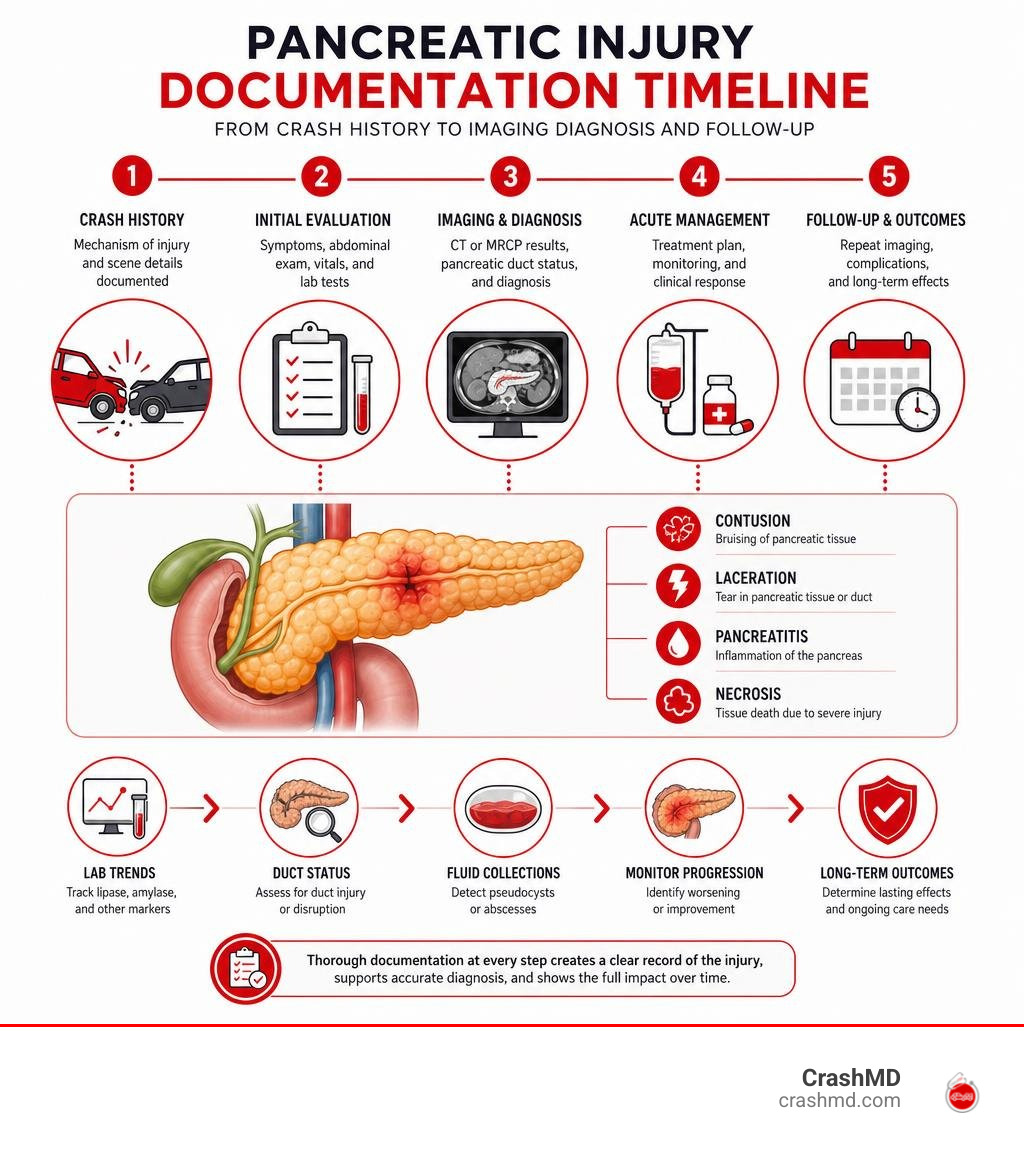
Quick look at medical records pancreas damage:
- the complete guide to how a pancreas injury affects the body
- pancreas injury survival guide severity symptoms
- a comprehensive guide to pancreas injury legal documentation
Understanding medical records pancreas damage is crucial for accurate diagnosis and treatment.
Understanding medical records pancreas damage is crucial for accurate diagnosis and treatment.
Diagnostic Standards and Medical Records Pancreas Damage Documentation

Accurate medical documentation requires strict alignment between clinical findings and diagnostic criteria. The pancreas is deeply nestled in the retroperitoneum, making clinical evaluation tricky. Digestive enzymes can activate prematurely within the gland, sparking self-digestion and tissue damage. Under the Revised Atlanta Classification, establishing a definitive diagnosis requires meeting at least two of three clinical benchmarks: classic epigastric abdominal pain radiating to the back, serum pancreatic enzyme elevation, and confirmatory cross-sectional imaging.
Reviewing ICD-10-CM clinical documentation guidance highlights that vague entries like “abdominal pain” or “pancreatic irritation” invite audit flags and billing denials. Every chart entry must clearly outline the underlying etiology, organ severity, and specific complications.
Differentiating Acute, Chronic, and Acute-on-Chronic Pancreatitis
Medical charts must distinguish between reversible acute inflammation and progressive chronic destruction:
- Acute Pancreatitis: Sudden inflammation triggered by gallstones, alcohol, trauma, or medications. About 80,000 cases occur in the United States annually, with roughly 20 percent categorized as severe. It is more common in men than women.
- Chronic Pancreatitis: Irreversible fibrosis, parenchymal calcification, and ductal dilatation. Chronic pancreatitis often develops in people between the ages of 30 and 40. In up to 70 percent of adult patients, chronic pancreatitis appears to be caused by alcoholism, while up to 30 percent of chronic pancreatitis cases have no apparent cause and are labeled idiopathic. Notably, up to 50 percent of chronic pancreatitis patients experience some pain relief when they stop drinking alcohol.
- Acute-on-Chronic Pancreatitis: An acute inflammatory flare superimposed on existing chronic structural damage.
When documenting acute-on-chronic episodes, providers must dual-code both the acute flare and the underlying chronic condition to capture clinical complexity.
Imaging Modalities, CT Severity Index, and Laboratory Biomarkers
Objective chart validation requires specific laboratory and radiological evidence:
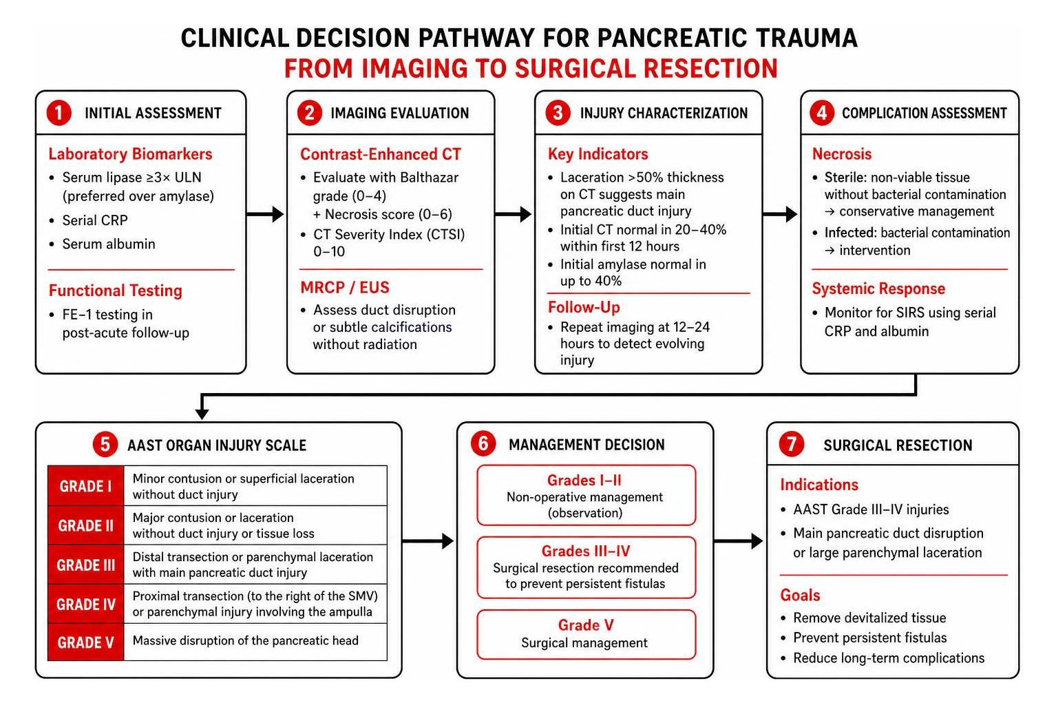
- Laboratory Biomarkers: Serum lipase is preferred over amylase because it stays elevated longer and offers higher pancreatic specificity. A level of at least three times the upper limit of normal satisfies the biochemical Atlanta criterion. Serial inflammatory markers, including C-reactive protein (CRP) and serum albumin, serve as prognostic indicators.
- Contrast-Enhanced CT & CTSI: A contrast-enhanced CT scan evaluated using the Balthazar grading scale (0 to 4 points) combined with pancreatic necrosis scores (0 to 6 points) creates the CT Severity Index (CTSI), ranging up to 10 points.
- Magnetic Resonance Cholangiopancreatography (MRCP): High-resolution MRCP or endoscopic ultrasound (EUS) is vital when assessing pancreatic duct disruption or subtle calcifications without radiation.
- Functional Testing: Post-acute follow-up evaluations utilize Fecal Elastase-1 (FE-1) testing to identify digestive enzyme deficits, supported by a study on acute pancreatic functional impairment.
Pancreatic Trauma, Necrosis, and Systemic Complication Profiles
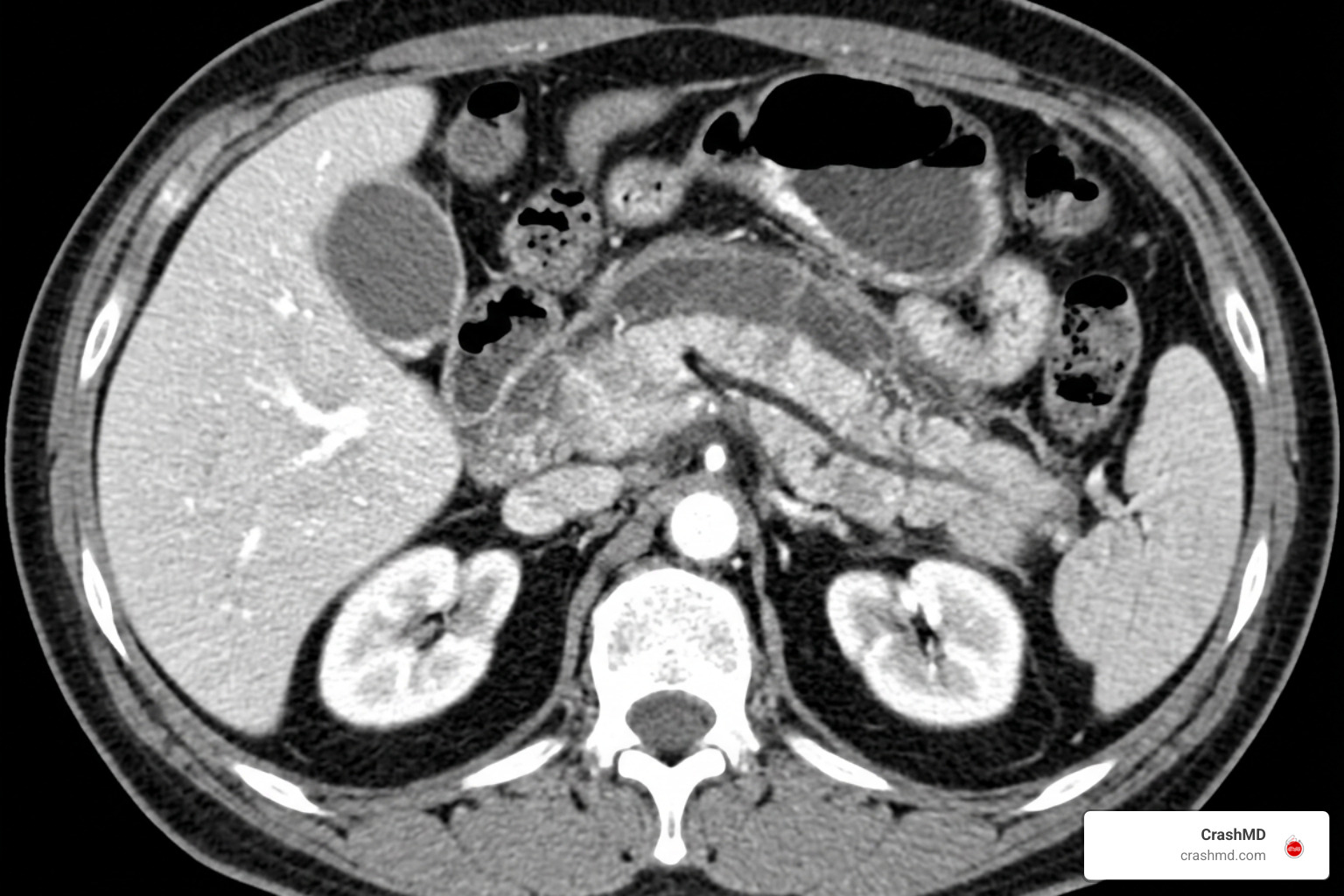
Pancreatic trauma accounts for up to 12 percent of abdominal injuries. The gland’s position across the lumbar spine leaves it vulnerable to compression from steering wheels or seatbelts during motor vehicle collisions. For detailed clinical management, see our guide on a comprehensive guide to ruptured pancreas severity and treatment.
Interpreting Medical Records Pancreas Damage from Blunt Force Trauma
Blunt abdominal trauma can easily be underdiagnosed early on because initial CT scans may appear normal in 20 to 40 percent of patients during the first 12 hours. Initial serum amylase can also be normal in up to 40 percent of cases. For this reason, follow-up imaging at 12 to 24 hours is essential to catch evolving tissue damage.
Trauma surgeons classify injury severity using the American Association for the Surgery of Trauma (AAST) Organ Injury Scale:
- Grade I: Minor contusion or superficial laceration without duct injury.
- Grade II: Major contusion or laceration without duct injury or tissue loss.
- Grade III: Distal transection or parenchymal laceration with main pancreatic duct injury.
- Grade IV: Proximal transection (to the right of the superior mesenteric vein) or parenchymal injury involving the ampulla.
- Grade V: Massive disruption of the pancreatic head.
A parenchymal laceration exceeding 50 percent of the gland’s thickness on CT strongly points to main pancreatic duct disruption. As highlighted in a clinical analysis of blunt pancreatic trauma and the EAST trauma surgical practice management guideline, Grade III and IV injuries usually require surgical resection rather than observation to prevent persistent fistulas. Detailed records are crucial for clinical and claim needs, as outlined in our a comprehensive guide to pancreas injury legal documentation.
Distinguishing Infected vs. Sterile Pancreatic Necrosis and SIRS
When digestive enzymes cut off local blood flow, non-viable tissue results in pancreatic necrosis. Charts must clearly differentiate between sterile and infected necrosis:
- Sterile Pancreatic Necrosis: Non-viable tissue without bacterial contamination, typically managed conservatively.
- Infected Pancreatic Necrosis: Bacterial invasion of dead tissue, typically appearing 2 to 3 weeks post-onset. Documentation requires culture confirmation, image-guided fine-needle aspiration, or visible retroperitoneal gas bubbles on CT.
This distinction is clinically and financially significant: sterile necrosis maps to MS-DRG 439 (Complication/Comorbidity tier), whereas infected necrosis elevates the case to MS-DRG 438 (Major Complication/Comorbidity tier).
Furthermore, non-infectious Systemic Inflammatory Response Syndrome (SIRS) must be documented using code R65.10 or R65.11, keeping it clearly distinct from infectious sepsis. A Bedside Index for Severity in Acute Pancreatitis (BISAP) score >= 3 is associated with 5–20% mortality risk in acute pancreatitis.
Long-Term Functional Complications and Secondary Diagnoses
Pancreatic trauma often causes lasting functional challenges. The table below outlines documentation requirements for secondary complications:
| Complication | Pathophysiology | Key Diagnostic Markers | ICD-10 Coding |
|---|---|---|---|
| Exocrine Pancreatic Insufficiency (EPI) | Loss of acinar tissue causing digestive enzyme deficits | Fecal Elastase-1 (<200 µg/g), steatorrhea, PEI-Q scores | K86.81 |
| Pancreatogenic Type 3c Diabetes | Destruction of endocrine islet beta cells | Fasting blood glucose, HbA1c elevation, loss of insulin/glucagon | E08.x series |
| Pancreatic Pseudocyst | Encapsulated fluid collection rich in pancreatic enzymes | Contrast CT, MRI/MRCP, abdominal ultrasound | K86.3 |
| Protein-Calorie Malnutrition | Malabsorption of fats and fat-soluble vitamins (A, D, E, K) | Serum prealbumin, albumin, unintended weight loss percentage | E43–E46 series |
Documenting Exocrine Panocrine Insufficiency and Malnutrition
Up to 26 percent of patients experience persistent exocrine dysfunction months after an initial severe pancreatic injury. When documenting Exocrine Pancreatic Insufficiency (EPI), medical records must detail oily, foul-smelling stools (steatorrhea), unintentional weight loss, and low fecal elastase-1 levels.
Documenting Pancreatic Enzyme Replacement Therapy (PERT) taken with meals is essential to capture ongoing care needs. For deeper insights into lasting post-accident effects, explore long-term pancreas injury effects.
Managing Pancreatogenic Type 3c Diabetes and Pseudocysts
Extensive injury to the islet cells impairs both insulin and glucagon regulation, leading to pancreatogenic diabetes (Type 3c). Coders must sequence the underlying pancreatic condition first, followed by the secondary diabetes code (E08 series).
If a pancreatic pseudocyst develops, records must detail its size, anatomical location, and interventions like endoscopic cystogastrostomy (CPT 43240) or percutaneous drainage.
Clinical Documentation Integrity, ICD-10-CM, and HCC Risk Adjustment
Accurate clinical documentation integrity (CDI) ensures health records reflect the actual severity of illness and support proper risk adjustment.
Resolving Coding Queries in Medical Records Pancreas Damage Charts
When chart notes are vague, CDI specialists send compliant, non-leading queries to attending physicians. Common query triggers include:
- Unspecified acute pancreatitis without etiology (e.g., clarifying post-traumatic K85.8x versus alcohol-induced K85.2x).
- Necrosis without confirmation of sterile versus infected status.
- Pancreatitis accompanied by transient organ failure lasting under 48 hours versus persistent organ failure exceeding 48 hours.
CMS-HCC V28 Mapping and Annual Documentation Requirements
Under the CMS-HCC (Hierarchical Condition Category) V28 risk adjustment model:
- Acute Pancreatitis: An episodic acute condition that does not map to a chronic HCC category.
- Chronic Pancreatitis (K86.0, K86.1): Maps directly to an HCC category, increasing the patient’s Risk Adjustment Factor (RAF) score.
To satisfy annual risk adjustment audits, medical providers must evaluate and document chronic pancreatic conditions at least once per calendar year using the MEAT framework (Monitor, Evaluate, Assess, Treat).
Frequently Asked Questions About Pancreatic Injury Documentation
How is traumatic pancreatic injury differentiated from disease-induced pancreatitis in health records?
Trauma records must document the physical mechanism (such as seatbelt sign or steering column impact), assign an AAST organ injury grade, note parenchymal laceration depth, and include external cause codes.
What laboratory and imaging thresholds confirm acute pancreatitis for medical coding?
Charts require at least two of the three Atlanta criteria: characteristic upper abdominal pain radiating to the back, serum lipase or amylase at least three times the upper limit of normal, and confirmatory cross-sectional CT or MRI findings.
How does documentation of pancreatic necrosis impact hospital MS-DRG assignment?
Uninfected or sterile necrosis maps to MS-DRG 439 (CC status). Documenting infected necrosis—supported by cultures, CT gas bubbles, or aspiration—elevates the case to MS-DRG 438 (MCC status), reflecting higher clinical complexity and resource utilization.
Conclusion
Thorough, objective medical chart documentation is essential for tracking recovery, supporting insurance claims, and guiding long-term care following a pancreatic injury. When clinical records precisely document diagnostic biomarkers, CT severity scores, duct integrity, and secondary functional deficits, patients receive the structured care they need.
At CrashMD, we help accident victims across Arizona, Nevada, Texas, Oklahoma, Southern California, and Northern California navigate their recovery journey with dedicated pain management and personalized treatment plans. If you or a loved one suffered a pancreas injury from a car accident, our medical team is here to support your physical healing and document your injuries thoroughly before taking legal steps.