

What Floating Ribs Pain Can Mean After an Injury
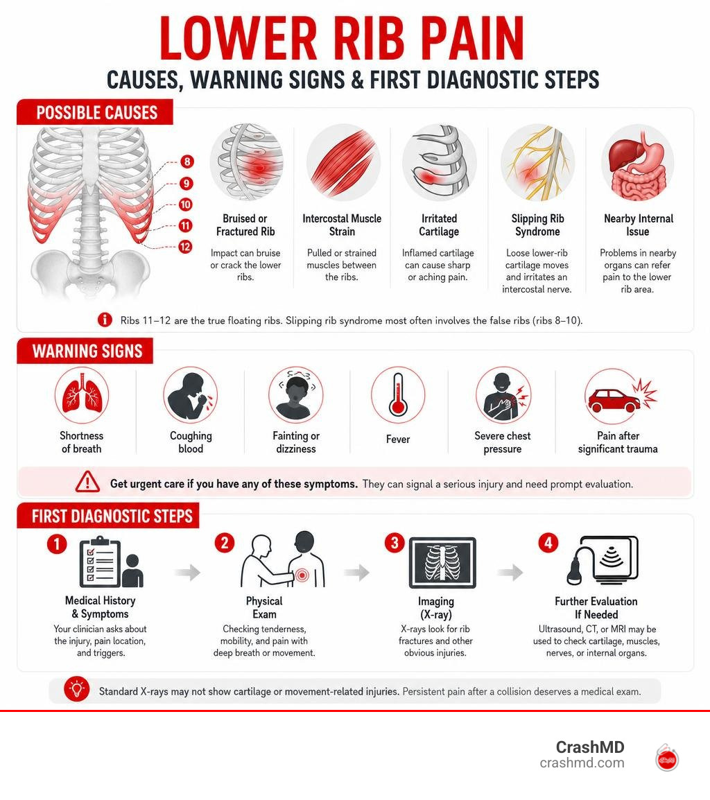
Floating ribs pain is discomfort near the lowest ribs, often felt in the side, back, flank, or upper abdomen. After a car accident or other impact, it may come from a bruised or fractured rib, an intercostal muscle strain, irritated cartilage, or a nearby internal problem. Sharp pain with breathing, coughing, twisting, or pressing the area is common.
A less obvious cause is slipping rib syndrome. Despite its nickname, this usually involves the false ribs (ribs 8-10), not the true floating ribs (ribs 11-12). Loose lower-rib cartilage can move and irritate an intercostal nerve, causing a click, pop, stabbing pain, or lingering ache.
Get urgent care for rib pain with shortness of breath, coughing blood, fainting, fever, severe chest pressure, or pain after significant trauma. Even without these warning signs, persistent pain after a collision deserves a medical exam because standard X-rays may not show cartilage or movement-related injuries.
I’m Dr. Adam Savage, and this guide explains what may be behind lower rib pain, how clinicians sort out the cause, and what relief and recovery can look like.
Floating ribs pain terms simplified:
Anatomy of the Lower Rib Cage: False Ribs vs. Floating Ribs
The human rib cage consists of 12 pairs of ribs attached posteriorly to the thoracic spine. However, their anterior attachments differ significantly, creating different structural vulnerabilities throughout the axial skeleton.
| Rib Type | Rib Numbers | Anterior Attachment | Primary Clinical Vulnerability |
|---|---|---|---|
| True Ribs | Ribs 1–7 | Direct costal cartilage to the sternum | Direct impact fractures |
| False Ribs | Ribs 8–10 | Indirect via shared costal cartilage arch | Hypermobility, Slipping Rib Syndrome |
| Floating Ribs | Ribs 11–12 | None (free-floating in muscle wall) | Rotational strain, 12th Rib Syndrome |
Anatomical Vulnerabilities in Lower Rib Structure
The 11th and 12th ribs are unique because they do not connect to the sternum or the costal cartilage arch. Instead, they embed directly into the musculature of the lateral abdominal wall, including the internal oblique, external oblique, and transversus abdominis muscles. While this structural configuration allows for necessary diaphragmatic expansion and trunk mobility, it leaves the surrounding intercostal nerves and costovertebral joints susceptible to rotational shearing forces and direct impact.
Because these floating ribs lack anterior tethering, high-energy impact forces or aggressive lateral flexion can disrupt their posterior articulations at the vertebral column. When the costovertebral ligaments become lax or sprained, the floating rib can pivot abnormally, irritating the neurovascular bundle running along its subcostal groove.
What Causes Floating Ribs Pain?
Pain in the lower thoracic region typically stems from several distinct mechanical and traumatic pathways:
- Mechanical Subluxation: Abnormal movement of loose rib tips or lax interchondral junctions.
- Nerve Impingement: Irritation or compression of the intercostal nerves traveling along the costal groove.
- Trauma: Seatbelt compression, direct blunt force, or steering wheel impact during motor vehicle collisions.
- Muscular Strain: Forceful coughing fits, heavy lifting, or sudden torso twisting that overloads the quadratus lumborum or intercostal musculature.
- Postural Strain: Prolonged poor seated biomechanics that compress the lower thoracic cage against the pelvic rim.
For comprehensive details on structural damage, review our detailed guide to rib cartilage injury or consult the MedlinePlus Medical Encyclopedia.
Identifying Common Causes of Floating Ribs Pain
Slipping Rib Syndrome and 12th Rib Syndrome
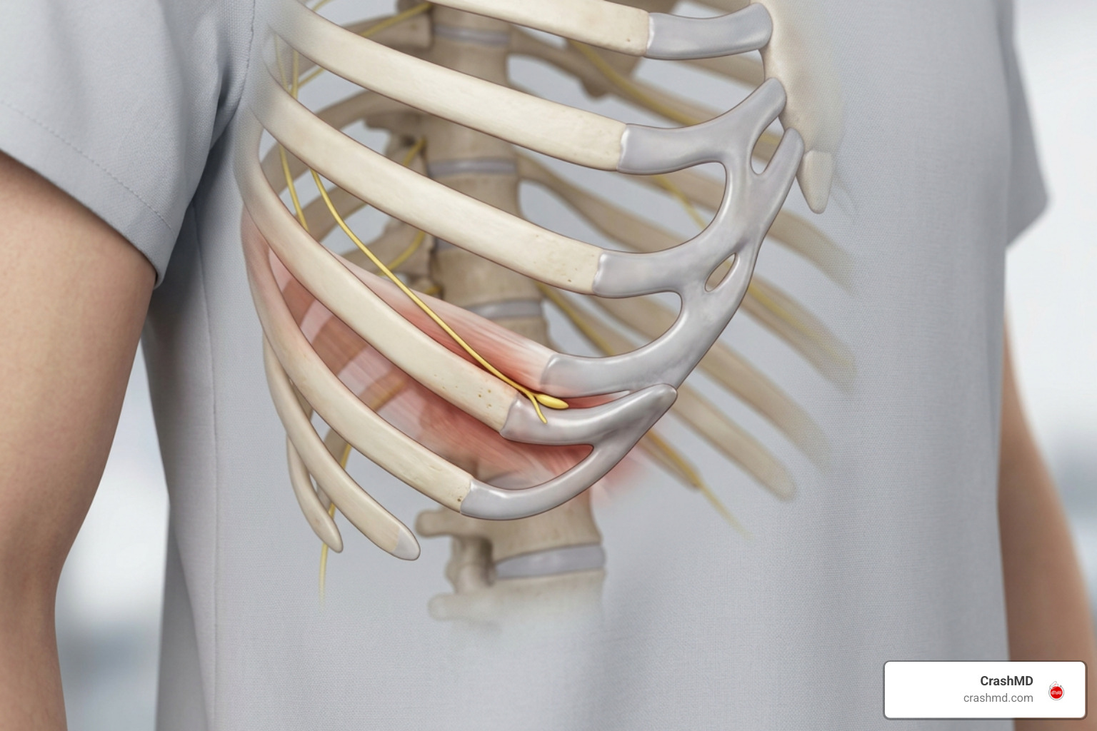
Slipping Rib Syndrome (also known as Cyriax syndrome) accounts for roughly 5% of musculoskeletal chest pain cases in primary care, with an estimated lifetime prevalence between 20% and 40%. It occurs when the weakened or torn fibrous cartilage band connecting ribs 8, 9, or 10 gives way. Without this stabilizing bridge, the affected rib tip slips and overrides the rib immediately above or below it, pinching the intercostal nerve.
A closely related disorder, 12th rib syndrome, involves hypermobility of the floating rib tips themselves. Because the 12th rib is unattached anteriorly, excessive mobility can compress the 12th thoracic (subcostal) or iliohypogastric nerve, irritating adjacent soft tissues and producing radiating pain in the flank, groin, buttocks, or lumbar region. As detailed in A Comprehensive Review of Slipping Rib Syndrome, this somatic nerve entrapment frequently mimics internal organ disorders such as kidney stones, gallbladder disease, or appendicitis.
Muscle Strains, Costochondritis, and Traumatic Fractures
Direct trauma from a collision frequently damages the soft tissues supporting the thoracic cage. Violent deceleration forces can cause severe strain to the intercostal muscles, latissimus dorsi, and serratus posterior inferior. For non-bony injuries, appropriate soft tissue injury treatment is critical to prevent chronic scarring and myofascial trigger points.
Costochondritis—inflammation of the costal cartilage junctions—can also manifest in the lower rib cage, producing continuous, burning anterior chest and rib pain that worsens with deep inspiration. Motor vehicle collisions often produce combined musculoskeletal injuries; understanding why your back hurts after a fender bender helps contextualize how spinal forces transfer across costovertebral joints to the lower ribs during sudden impact.
Clinical Diagnosis: How Doctors Evaluate Lower Rib Cage Pain
The Physical Exam and the Hooking Maneuver
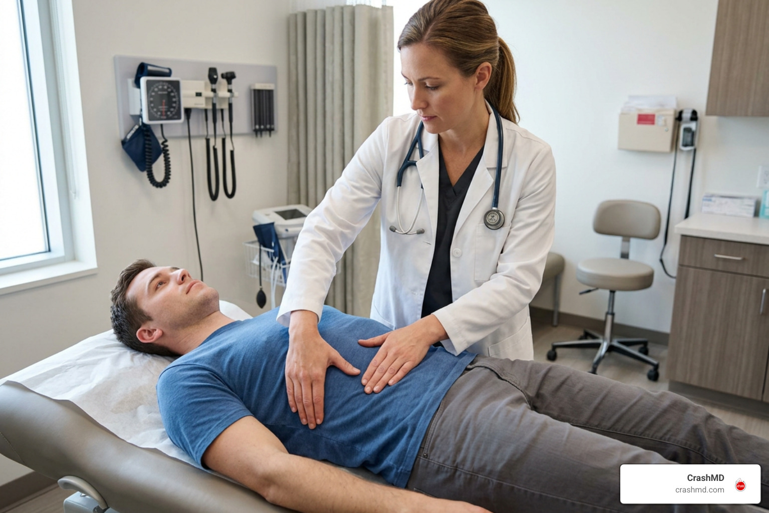
Because costal cartilage is radiolucent (invisible on standard X-rays), a thorough physical evaluation is the cornerstone of an accurate diagnosis. Skilled clinicians systematically palpate the costal margins, assessing for point tenderness, intercostal muscle spasms, and structural asymmetry along the lower rib cage.
Providers frequently use the hooking maneuver, placing their fingers under the lower anterior costal margin and gently pulling anteriorly and superiorly. A positive test reproduces the patient’s characteristic sharp, radiating pain and may elicit an audible or palpable click as the hypermobile rib subluxates over the adjacent rib border. Identifying this localized click allows clinicians to differentiate cartilage instability from visceral pathology.
Dynamic Ultrasound vs. Standard Static Imaging
Standard static X-rays and CT scans are typically performed with the patient lying supine—a position that offloads the rib cage and reduces subluxation, frequently yielding false-negative results.
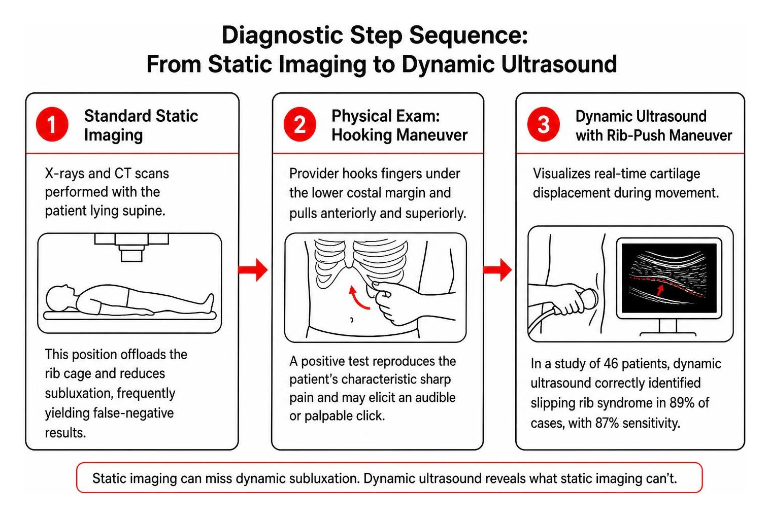
Dynamic high-resolution ultrasound has emerged as the premier imaging modality for evaluating lower rib cage hypermobility and cartilage disruptions. In a study of 46 patients, dynamic ultrasound correctly identified slipping rib syndrome in 89% of cases. Using the targeted “rib-push maneuver,” dynamic ultrasound achieves 87% sensitivity by visualizing real-time cartilage displacement during movement, deep inspiration, and targeted probe pressure, clearly identifying the mechanical impingement that static scans miss.
Comprehensive Treatment and Management Strategies
We employ a stepped-care model starting with conservative measures before evaluating interventional procedures, tailoring each intervention to the specific cause of pain.
Conservative Care and Interventional Nerve Blocks
Initial management centers on calming local inflammation and protecting the nerve from recurring mechanical irritation:
- Thermal Therapy: Ice application during acute flares to numb sharp pain, followed by heat to relax guarding thoracic muscles.
- Pharmacotherapy: Scheduled NSAIDs, acetaminophen, muscle relaxants, or topical lidocaine patches applied directly over the costal margin.
- Activity Modification: Avoiding high-torque trunk rotation, sudden bending, and heavy lifting while the cartilage and intercostal fibers rest.
- Targeted Physical Therapy: Core stabilization, postural re-education, and diaphragmatic breathing training to offload tension from the lower rib cage.
- Intercostal Nerve Blocks: Injections of a local anesthetic and corticosteroid directly beneath the affected rib provide both diagnostic confirmation and therapeutic relief by breaking the chronic pain cycle.
Surgical Options: Cartilage Resection and Stabilization
For refractory cases where conservative therapies fail to provide durable relief, thoracic specialists may evaluate surgical interventions. Modern surgical techniques include minimally invasive costal cartilage excision or vertical rib stabilization using bioabsorbable plates and permanent sutures to secure the hypermobile rib tips. In a cohort of 30 patients undergoing surgical repair, 44% achieved complete pain resolution postoperatively, and all experienced significant improvement. Clinical protocols published by the Cleveland Clinic highlight that stabilizing the hypermobile margin prevents recurrent nerve impingement and restores functional rib cage integrity.
Recovery Outlook and Long-Term Prevention
Healing Timelines and Managing Floating Ribs Pain Flare-Ups
Understanding expected healing timelines helps set realistic milestones and prevents premature return to high-risk physical activities:
- Intercostal Muscle Strains: Typically resolve in 1 to 3 weeks with adequate rest and gradual stretching.
- Bruised or Fractured Ribs: Generally require 4 to 6 weeks for primary bony healing, though localized tenderness may persist slightly longer.
- Cartilage and Nerve Injuries: Cartilage has a limited blood supply, meaning hypermobility and intercostal neuralgia may cause intermittent flare-ups for several months.
Because thoracic symptoms can evolve over days or weeks following impact, monitoring delayed symptoms after a car accident ensures subtle cartilage displacement, costovertebral joint sprains, or nerve entrapment is caught early before progressing into chronic neuropathic pain.
Seeking Medical Care After Trauma
Following an auto collision, comprehensive clinical evaluation and detailed medical documentation are essential to support both physical recovery and personal injury documentation. Proper injury charting links your symptoms directly to the traumatic incident, protecting your health and legal rights. If you have sustained impact trauma, review our clinical protocols on rib cartilage injury after car accident.
Frequently Asked Questions About Floating Rib Pain
Can stress and anxiety worsen lower rib cage pain?
Yes. Stress increases muscular tension throughout the thoracic wall, leading to shallow chest breathing and intercostal muscle spasms that lower pain thresholds and aggravate irritated nerves.
What are the emergency red flags for floating rib injuries?
Seek immediate emergency medical evaluation if lower rib pain is accompanied by shortness of breath, coughing up blood, sudden dizziness, high fever, or abdominal rigidity.
How does slipping rib syndrome differ from a cracked rib?
A cracked rib is a structural fracture of the cortical bone, visible on detailed radiographic imaging, which heals as bone knits over 4 to 6 weeks. Slipping rib syndrome is a mechanical hypermobility of the costal cartilage that slips over adjacent ribs, repeatedly pinching the intercostal nerve.
Conclusion
Lower rib pain requires careful clinical distinction between bone fractures, muscular strains, and cartilage hypermobility like slipping rib syndrome. At CrashMD, we provide comprehensive injury evaluation, advanced pain management, and objective medical documentation for accident victims across Arizona, Nevada, Texas, Oklahoma, Southern California, and Northern California, ensuring your recovery is supported every step of the way.