

Understanding Rib Cartilage Injury: Anatomy, Causes, and Types
A rib cartilage injury is a tear, separation, or fracture of the flexible hyaline cartilage that connects your ribs to your breastbone. Recovery typically follows a phased course: protecting the chest for the first few weeks, gradually restoring movement over the next month or two, and returning to full activity only after pain-free breathing and rotation are restored. Most injuries heal without surgery, but accurate imaging and structured rehabilitation are essential because standard X-rays cannot see cartilage damage.
The human rib cage consists of 12 pairs of ribs, but they do not all attach directly to the breastbone (sternum) as solid bone. Instead, the anterior portion of your rib cage relies on costal cartilage—a tough, elastic form of hyaline cartilage. This cartilage creates critical structural bridges at two main points:
- Chondrosternal joints: Where the cartilage connects directly to the sternum.
- Costochondral junctions: Where the bony rib transitions into the flexible cartilage segment.
True ribs (ribs 1–7) attach directly to the sternum via their own costal cartilage. False ribs (ribs 8–10) connect to the cartilage of the rib above them, while floating ribs (ribs 11–12) end freely in the abdominal wall. This cartilaginous network provides the thoracic elasticity required to expand and contract thousands of times a day during respiration.
A sudden blunt trauma—such as the rapid deceleration against a seatbelt during a motor vehicle collision, a fall, or an intense athletic collision—can exceed the physical threshold of this flexible tissue, leading to a severe radiological reference on costal cartilage injuries.
| Feature | Bony Rib Fracture | Costal Cartilage Injury |
|---|---|---|
| Tissue Type | Vascularized bone | Avascular hyaline cartilage |
| X-Ray Detection | Readily visible (50–75% sensitivity) | Completely invisible (radiolucent) unless heavily calcified |
| Primary Location | Posterior or lateral rib cage | Anterior chest wall along the sternal border |
| Blood Supply | Rich periosteal vascular network | Minimal intrinsic blood flow (relies on perichondrium) |
| Average Healing Time | 6 to 8 weeks | 6 weeks to 6+ months |
| Common Sensation | Deep ache, grinding crepitus | Sharp catching pain, audible popping/clicking |
Costochondral Separation vs. Cartilage Fracture
While both conditions stem from thoracic trauma, their mechanics differ:
- Costochondral Separation: The cartilage tears completely away from the bony rib shaft at the junction. This often occurs from violent rotational forces, such as sudden torso twisting while braced against a steering wheel, or heavy contact in collision sports.
- Costal Cartilage Fracture: A distinct crack, fissure, or complete structural break within the cartilage body itself. This injury usually stems from direct, localized blunt impact compressing the sternum and anterior chest wall.
Understanding these distinctions is essential for planning long-term care. You can explore our detailed guide to rib cartilage injury to see how specific accident vectors alter the damage pattern.
Differentiating Traumatic Injury from Costochondritis
Chest wall pain is one of the most common reasons patients present to acute care clinics. In fact, noncardiac, musculoskeletal issues account for 33% to 47% of outpatient chest pain visits and up to 45% of noncardiac emergency room evaluations (Schumann et al., 2024).
Patients frequently ask whether their injury is a traumatic tear or costochondritis:
- Costochondritis: A benign, inflammatory condition affecting the costochondral or chondrosternal joints without traumatic structural disruption. It causes localized tenderness—most commonly across the 2nd through 5th ribs—without external swelling (Schumann et al., 2024). Over 90% of cases improve within 3 to 4 weeks with conservative care (Schumann et al., 2024). A related inflammatory condition, Tietze syndrome, presents similarly but includes visible swelling, warmth, and erythema.
- Traumatic Cartilage Fractures: These involve physical disruption of the cartilage matrix, sharp mechanical pain during movement, occasional popping sensations, and longer healing trajectories that require objective diagnostic documentation. Reviewing clinical research on costochondritis helps clinicians accurately separate inflammatory conditions from mechanical trauma.
Diagnostic Protocols: Imaging and Clinical Evaluation
Diagnosing anterior chest trauma begins with a meticulous physical examination. Clinicians use specific palpation maneuvers to isolate costochondral joints:
- Crowing Rooster Test: The examiner stands behind the patient, placing hands under the rib cage while the patient gently extends their neck and torso backward. This stretches the anterior chest wall to reproduce cartilage discomfort.
- Horizontal Arm Flexion Test: The patient’s arm is pulled across the chest while pressure is applied to the joint to evaluate mechanical joint stress.
Diagnosing a Rib Cartilage Injury: Why Standard X-Rays Fail
Standard chest radiographs (X-rays) are designed to absorb ionizing radiation through dense, calcium-rich bone. Hyaline cartilage is radiolucent—meaning X-rays pass straight through it without registering an image.
Unless a patient is elderly and has developed extensive, age-related physiological cartilage calcification, standard X-rays will appear completely normal. This can leave injured patients feeling invalidated when they are discharged with “clear” films despite debilitating pain. If you find yourself in this situation, our analysis on why your chest still aches weeks after a sudden collision explains why normal X-rays frequently miss soft tissue disruptions.
Dynamic Ultrasound, CT, and MRI Modalities
To confirm an occult costal cartilage disruption, advanced imaging is required:
- Dynamic High-Resolution Ultrasound: Ultrasound is non-invasive and allows for “sonar palpation.” The sonographer places the transducer over the tender area and asks the patient to twist or breathe deeply, allowing real-time assessment of cartilage separation, step-off deformities, and joint subluxation.
- Computed Tomography (CT) with 3D Reconstruction: Essential in multi-trauma settings to exclude organ damage, pneumothorax, and occult bony fractures (Kuo & Kim, 2023). Traumatic healing manifests as distinct vertical calcifications along fracture lines, unlike normal patchy physiological calcification patterns.
- Magnetic Resonance Imaging (MRI): T2 fat-saturated and TIRM sequences highlight bone marrow edema, perichondrial disruption, and regional soft tissue inflammation that other tests miss.
A prospective study on costal cartilage fractures in polytrauma showed that up to 38.1% of patients exhibit persistent edema on T2 MRI years after the initial event, highlighting why comprehensive cross-sectional imaging is critical for accurate diagnosis and legal documentation.
Treatment and Pain Management Protocols
Treating anterior chest wall injuries requires a balanced approach: managing acute pain effectively while protecting pulmonary function.
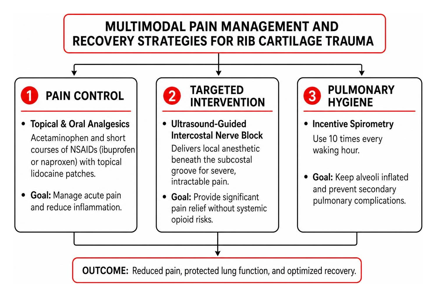
Aggressive pain management prevents respiratory splinting (shallow breathing due to pain). We recommend a multi-step protocol:
- Topical and Oral Analgesics: First-line care includes acetaminophen and short courses of Non-Steroidal Anti-Inflammatory Drugs (NSAIDs) like ibuprofen or naproxen, paired with topical lidocaine patches.
- Targeted Interventional Procedures: For severe, intractable pain, an ultrasound-guided intercostal nerve block delivers local anesthetic directly beneath the subcostal groove, providing significant pain relief without systemic opioid risks.
- Pulmonary Hygiene: Utilizing an incentive spirometer 10 times every waking hour ensures your alveoli remain inflated, preventing secondary pulmonary complications.
If you are dealing with acute post-impact soreness, our practical guide to immediate broken rib pain relief outlines safe home interventions.
Conservative Care and Why Chest Binding is Contraindicated
A common historical mistake was wrapping a tight elastic bandage or binder around a patient’s chest to immobilize the ribs.
Modern clinical guidelines strictly contraindicate chest binding.
Constricting the rib cage prevents deep inspiration and restricts lung expansion. This hypoventilation leads directly to pulmonary atelectasis (the collapse of small airways) and dramatically spikes the risk of pneumonia (Kuo & Kim, 2023). Modern management focuses on unrestrained breathing supported by targeted cold therapy and safe analgesia, as outlined in this clinical overview of rib trauma management.
Indications for Surgical Stabilization and Plating
Most cartilage injuries resolve with non-operative care. However, surgical stabilization via open reduction and internal fixation (ORIF) may be required when:
- There is an unstable flail chest segment (three or more consecutive ribs broken in two or more places, causing paradoxical chest motion) (Kuo & Kim, 2023).
- The patient fails to wean from mechanical ventilation due to chest wall deformity.
- Chronic, painful non-union or gross structural dislocation persists for months.
Surgeons use low-profile titanium plates anchored by locking screws across the costal cartilage and sternum. Reviewing advanced surgical techniques for cartilaginous anterior chest wall repair shows that placing at least three bicortical locking screws on each side of the fracture provides excellent biomechanical stability and long-term symptom relief.
Phased Rehabilitation and Healing Timeline
Because hyaline cartilage is avascular, chondrocyte repair occurs slowly through peripheral diffusion from the perichondrium. Rather than generating rapid bony callus, the body bridges cartilage breaks with dense fibrocartilaginous tissue.

Phased Timeline for Rib Cartilage Injury Recovery
- Acute Protective Phase (Weeks 1 to 3): Focus on resting, controlling inflammation, gentle walking, and diligent incentive spirometry. Avoid all lifting, twisting, and abrupt pushing or pulling.
- Subacute Healing Phase (Weeks 4 to 8): Pain during resting respiration should subside. Begin gentle posture retraining, light scapular retractions, and active thoracic extension.
- Remodeling and Functional Return (Weeks 9 to 16+): Gradually introduce progressive resistance training, core stabilization, and sport-specific rotational movements once cleared by a clinician.
For a detailed phase-by-phase breakdown, consult the ultimate guide to rib cartilage injury recovery timeline along with this symptoms and recovery overview.
Long-Term Complications and Preventing Chronic Instability
Long-term follow-up studies in blunt polytrauma reveal that up to 23.8% of patients experience progressive dislocation over time, with nearly 20% suffering lingering discomfort, chronic cough, and popping sensations years down the road.
Unstable lower rib cartilage can also lead to slipping rib syndrome, where hypermobile costal margins irritate the adjacent intercostal nerves. If your injury was caused by a motor vehicle collision, having an expert evaluation for a rib cartilage injury after a car accident ensures your structural damage is medically stabilized and fully documented for personal injury insurance claims.
Frequently Asked Questions About Rib Cartilage Injuries
Can fractured rib cartilage ever fully heal on its own?
Yes, but it typically heals through fibrocartilaginous scar tissue rather than regenerating pristine hyaline cartilage. While this scar bridge stabilizes the joint and resolves pain in most cases, the structural area may remain slightly less pliable than before the trauma.
How do I know if my rib pain is from cartilage or bone?
Bone fractures typically create pinpoint pain along the sides or back of the rib cage. Cartilage damage is characterized by sharp tenderness along the anterior sternal border, often accompanied by an audible click or slipping sensation during deep rotational twists. Definitive diagnosis requires dynamic ultrasound, CT, or MRI.
When is it safe to return to contact sports or heavy lifting?
You should return to contact sports only when you have achieved 100% pain-free thoracic rotation, can take a maximal deep breath without discomfort, and exhibit no tenderness on palpation. Athletes returning to high-impact environments should wear fitted, protective chest padding to avoid re-injury.
Conclusion
Costal cartilage injuries are painful, easily overlooked trauma conditions that require a clear diagnostic strategy. Because plain X-rays cannot visualize damaged hyaline cartilage, relying on dynamic ultrasound, high-resolution CT, and MRI is essential to uncover hidden injuries, prevent respiratory complications, and guide appropriate rehabilitation.
At CrashMD, we guide accident victims across Arizona, Nevada, Texas, Oklahoma, and California through every stage of recovery. We deliver specialized post-accident medical evaluations, multimodal pain management, and objective injury documentation to protect your health and support your legal claims before settlement negotiations begin.
If you are experiencing lingering anterior chest pain or clicking following a collision, schedule your comprehensive medical evaluation with our team today.
Medical Citations
Jessica A. Schumann, Tanuj Sood, John J. Parente. “Costochondritis.” StatPearls Publishing, 2024. NBK532931. Kevin Kuo, Amie M. Kim. “Rib Fracture.” StatPearls Publishing, 2023. NBK541020.