

Understanding Rib Anatomy and Cartilage Injury Symptoms
To understand how to heal rib cartilage injury, it helps to know how the human ribcage is constructed and how its structural components function together under physical stress. The human thoracic skeleton features 24 individual ribs (12 pairs) composed of roughly two-thirds solid bone and one-third dense hyaline cartilage, known as costal cartilage.
- True Ribs (1–7): Attach directly to the central breastbone (sternum) via individual bars of costal cartilage, providing vital anterior support for the chest wall.
- False Ribs (8–10): Attach indirectly to the sternum by joining the primary costal cartilage of the 7th rib, forming a flexible cartilaginous arch along the lower ribcage.
- Floating Ribs (11–12): Attach only to the thoracic vertebrae in the spine, ending freely within the surrounding abdominal and back musculature.
Costal cartilage gives your chest wall the flexibility needed to expand and contract smoothly during everyday breathing. However, when physical trauma occurs—such as during motor vehicle collisions or high-impact contact sports—this resilient tissue can bruise, tear, or completely detach from the bony rib shaft (a condition known medically as costochondral separation).
Symptoms of a cartilage injury include intense localized sternal tenderness, sharp chest pain during deep inhalation, and agonizing discomfort while coughing, laughing, or sneezing. You might even hear or feel a distinct crunching or popping sound (crepitus) during torso movements or rotational twisting. Because rib cartilage lacks the dense mineral structure of bone, healing requires targeted care. For a deeper breakdown of these complex structural issues, review our Detailed Guide to Rib Cartilage Injury.
Common Causes of Costal Cartilage Damage
Cartilage injuries stem from both sudden high-velocity physical impacts and severe, repetitive mechanical stresses:
- Blunt Force Trauma: Steering wheel collision, airbag deployment, or heavy seatbelt compression during auto accidents represents a leading cause of costal cartilage rupture and sternal separation.
- Contact Sports: Direct blows, tackles, or blunt strikes in martial arts, rugby, lacrosse, or football frequently compress and twist the lower chest wall.
- Severe Coughing Fits: Violent, prolonged coughing spells caused by respiratory infections exert intense internal thoracic pressure, straining chest tissue and tearing delicate cartilage fibers away from the bone.
- Repetitive Rotational Strain: Sudden athletic movements like golf swings, tennis serves, or rowing create torque and rotational stress that pull forcefully on intercostal joints.
Diagnostic Techniques and Clinical Evaluation
Diagnosing a rib cartilage issue begins with a comprehensive clinical examination. A trained medical provider conducts careful localized palpation and specialized procedures, such as the hooking maneuver (gently pulling the lower rib cage forward to evaluate joint hypermobility and lower rib instability).
Standard emergency room X-rays check for acute bone fractures but cannot directly visualize soft tissue structures like cartilage. If severe, sharp chest pain continues despite completely clear X-ray findings, expert clinical guidance recommends advanced imaging modalities tailored for soft-tissue assessment:
- CT Scans: Highly effective for revealing subtle non-displaced cartilage breaks, sternal joint shifts, and complex chest wall micro-fractures.
- MRI Scans: Recognized as the gold standard imaging modality for mapping soft tissue tears, localized intercostal inflammation, bone marrow edema, and joint fluid accumulation.
How to Heal Rib Cartilage Injury: Phase 1 Protocols (Days 1–14)
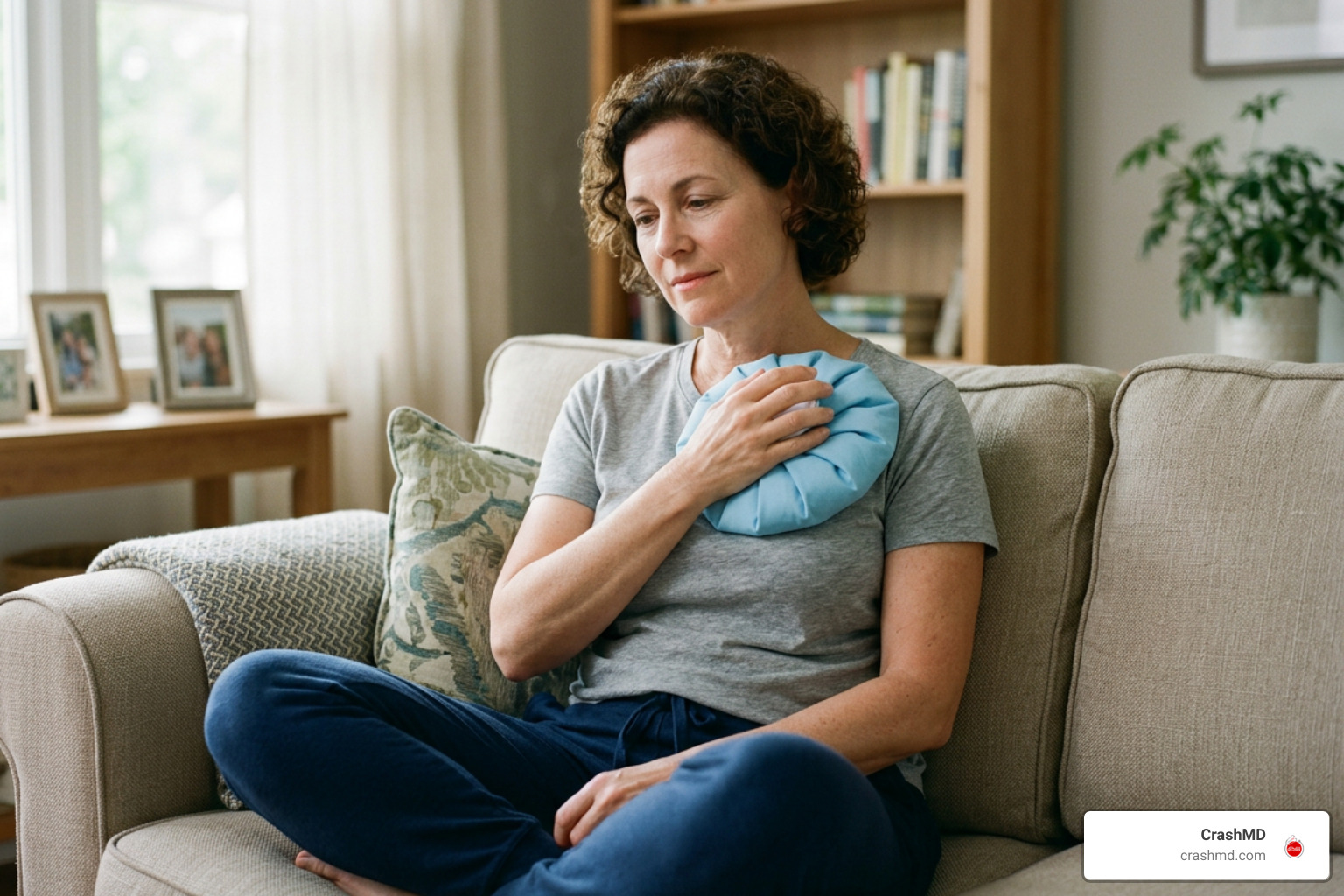
The primary goal during the initial two weeks of recovery is actively managing acute inflammation while maintaining normal lung expansion. After a traumatic impact damages your costal tissue, proper early stabilization is vital for setting the stage for long-term recovery. To better understand how soft tissue heals after severe physical impact, see our guide on Sprains, Strains, and Beyond: Understanding Soft Tissue Injuries.
Step-by-Step Guide on How to Heal Rib Cartilage Injury at Home
- Apply Cold Therapy Early: Place ice packs wrapped in a thin cloth over the tender site for 15–20 minutes at a time, every 2 to 3 hours during the first 48 hours to restrict blood vessel dilation and control localized swelling.
- Utilize Anti-Inflammatory Support: Take over-the-counter NSAIDs like ibuprofen or naproxen as recommended by your physician to lower systemic inflammation and calm tender intercostal nerve pathways.
- Practice Correct Sleep Positioning: Sleep on your back or on the uninjured side, using extra supportive pillows to keep your torso aligned and prevent nocturnal pressure on damaged costal tissue.
- Maintain Upright Posture: Avoid slouching, rounding your shoulders, or hunching over, which compresses inflamed costal joints and aggravates ongoing pain.
- Skip Tight Chest Binders: Restricting chest wall movement with rigid wraps or tight compression bands reduces breathing volume and significantly increases the risk of lung tissue collapse and pulmonary infections.
- Prevent Respiratory Illness: Avoid contact with sick individuals and practice proper hygiene to minimize the risk of catching a cold or infection that triggers painful coughing or sneezing fits.
Pain Management and Reducing Inflammation
Because broken ribs or torn rib cartilage cannot be stabilized in a traditional orthopedic cast, pain management is critical to allow adequate chest expansion during breathing. Effective clinical and home strategies include:
- Topical Analgesics: Over-the-counter or prescription lidocaine patches and anti-inflammatory gels applied near the injury site offer targeted pain control without gastrointestinal side effects.
- Therapeutic Heat: Transitioning to gentle heat packs after the initial 48-hour acute phase helps relax tight, spasming intercostal muscles and promotes localized blood circulation.
- Intercostal Nerve Blocks: Targeted clinical injections performed by a pain specialist that temporarily block local nerve impulses, granting relief so you can breathe deeply without excruciating discomfort.
Phase 2 Recovery: Rehabilitative Exercises and Active Healing (Days 15–30)
During days 15 through 30, your body transitions from acute inflammation control to active matrix repair, laying down fibrous scar tissue across the injured cartilaginous region. Transitioning into gentle, progressive rehabilitation prevents long-term thoracic stiffness while building dynamic stability around affected rib joints.
Nutrition and Exercise Strategies on How to Heal Rib Cartilage Injury
Because cartilage is an avascular structure with limited direct internal blood supply, oxygen and crucial nutrients reach the tissue slowly through joint fluid diffusion. Target these gentle movement patterns to stimulate fluid movement without straining healing fibers:
- Diaphragmatic Breathing: Lie flat on your back and focus on expanding your lower abdomen rather than lifting your ribcage, taking slow, deep, controlled breaths for 5 minutes twice daily to preserve baseline lung capacity.
- Transverse Abdominis Engagement: Practice mild core bracing (gently drawing your navel toward your spine) to build a natural internal corset that stabilizes the lower ribs during daily activities.
- Doorway Chest Stretch: Stand in a doorway, place your forearms lightly on the doorframe, and step forward gently until you feel a mild stretch across your chest, relieving pectoral tension that pulls on delicate costal cartilage.
Supplementing your daily diet with collagen peptides, Vitamin C, zinc, and omega-3 fatty acids supports extracellular matrix synthesis and tissue remodeling. To learn more about supportive dietary protocols and nutritional strategies, explore The Best Cartilage Repair Supplements to Keep You Moving.
Preventing Long-Term Complications and Chronic Costochondritis
Restricting your breathing due to sharp chest pain can lead to secondary pulmonary complications, including partial lung collapse (atelectasis) and pneumonia. Regularly expanding your lungs with gentle deep breathing exercises is essential during every phase of recovery.
Without appropriate initial care and structured rehabilitation, torn costal cartilage can fail to unite, forming a painful nonunion or progressing into chronic costochondritis—a persistent condition characterized by long-term inflammation of the sternocostal joints. In complex cases marked by severe mechanical instability, clicking, or persistent nonunion, specialized minor surgical procedures (such as internal thread stabilization or rib plate fixation) may be evaluated by an orthopedic surgeon.
For complete details on extended recovery timelines and long-term milestones, consult The Ultimate Guide to Rib Cartilage Injury Recovery Timeline.
Frequently Asked Questions about Rib Cartilage Healing
How long does it take for a rib cartilage injury to heal completely?
Most patients experience notable symptomatic improvement within 4 to 6 weeks under conservative physical care. However, because costal cartilage has a limited blood supply, complete structural tissue healing generally takes 6 to 12 weeks. Severe cartilage tears or complete costochondral separations can require up to 6 months for full tissue remodeling and joint stabilization.
How do I know if my rib cartilage injury is serious enough for surgery?
Surgical intervention is rarely required for rib cartilage damage. Conservative care successfully manages roughly 80% to 90% of hypermobile or slipping rib issues. Surgery (such as costal cartilage reduction or stabilization procedures like the Hansen method) is typically reserved for severe structural displacement, nerve entrapment, or chronic nonunion lasting months despite intensive physical therapy.
What is the difference between costochondritis and a torn rib cartilage?
Costochondritis refers to localized inflammation of the cartilage connecting the ribs to the sternum, often occurring without physical tissue rupture or macro-trauma. A torn rib cartilage involves an actual structural rupture, tear, or separation of the cartilage tissue caused by blunt trauma, severe impact, or sudden extreme rotational forces.
Conclusion: Achieving Complete Recovery and Next Steps
Reaching the 30-day mark in your recovery signals an important functional transition. By combining early ice therapy, active pain management, diaphragmatic breathing exercises, and gradual core stabilization, you lay the necessary structural foundation for complete chest wall healing and a safe return to regular physical activities.
If your rib injury occurred during a motor vehicle collision, proper medical documentation is essential for both your physical health and any related legal claims. At CrashMD, our clinical team supports accident victims across Arizona, Nevada, Texas, Oklahoma, and California through every step of physical recovery. We ensure your chest trauma is accurately evaluated with appropriate imaging, thoroughly documented by legal-medical standards, and properly treated before any legal claims are finalized.
To learn more about accident-related chest wall care and specialized recovery strategies, visit our comprehensive resource on Rib Cartilage Injury After Car Accident.