

What Is a Rib Cartilage Injury and Why It Matters After Trauma
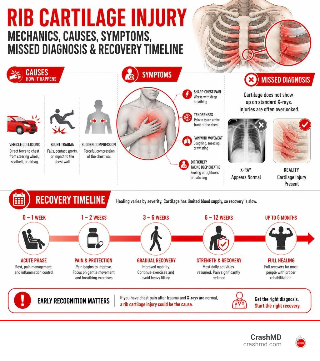
A cartilage injury in the rib is one of the most painful and most commonly missed chest injuries after a car accident or blunt trauma. Here is what you need to know at a glance:
- What it is: Damage to the costal cartilage — the tough, flexible tissue connecting your ribs to your breastbone
- Why it’s missed: Cartilage does not show up on standard X-rays, so many patients are sent home with a “normal” result despite a real injury
- Key symptoms: Sharp chest pain with breathing, tenderness at the front of the chest, and pain that worsens with movement or coughing
- Healing time: Typically 6 to 12 weeks, but can take up to 6 months due to cartilage’s limited blood supply
- Treatment: Most cases are managed with rest, NSAIDs, and breathing exercises — severe cases may require surgical stabilization
Rib cartilage injuries are estimated to occur in 20 to 42% of patients who suffer high-energy blunt chest trauma, yet they are frequently overlooked because standard imaging cannot detect them. That means thousands of accident victims leave the emergency room with unexplained, debilitating chest pain and no clear diagnosis.
If you are struggling to breathe deeply, feeling sharp pain in your chest wall after a collision, or were told your X-rays looked “fine” despite ongoing pain — you are not imagining it.
I’m Dr. Adam Savage, and in this guide I’ll walk you through everything you need to understand about rib cartilage injuries — from anatomy and diagnosis to treatment and recovery — so you can get the answers and care you deserve.
Understanding Rib Cage Anatomy and Types of Rib Cartilage Injury
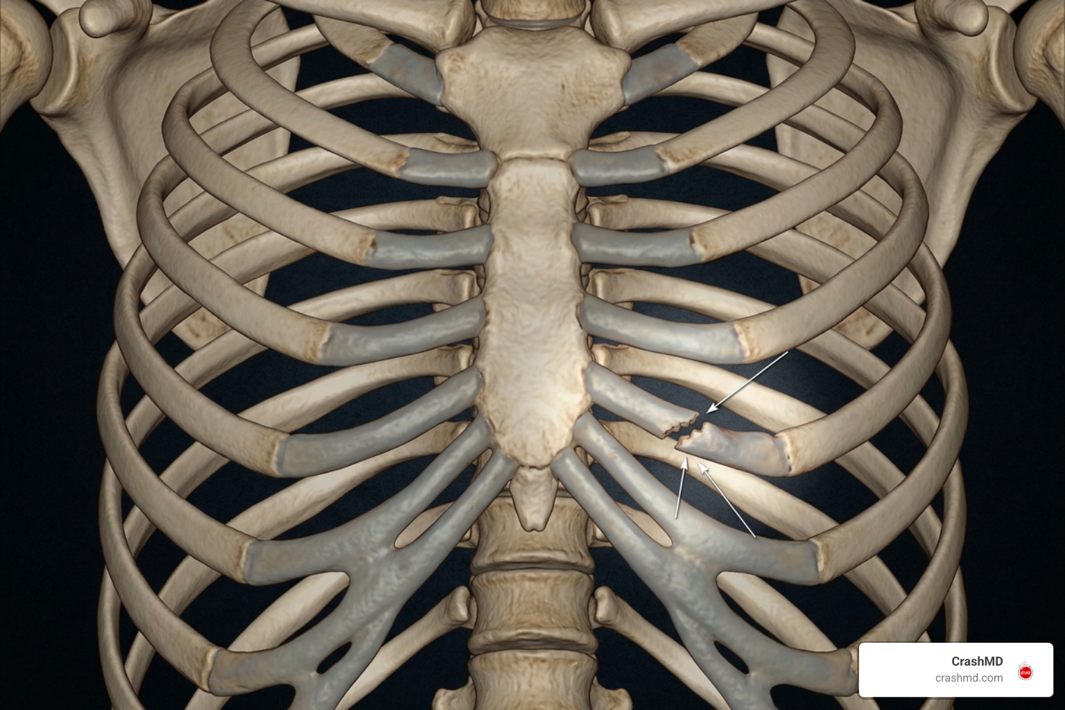
To understand how a cartilage injury rib trauma occurs, we first have to look at how the human chest wall is built. The thoracic cage consists of 24 curved ribs arranged in 12 pairs. However, bone doesn’t attach directly to your breastbone (sternum) across the entire front of your chest. Instead, hyaline cartilage—known as costal cartilage—acts as a flexible bridge between your rigid bone and sternum.
This setup is classified into three anatomical zones:
- True Ribs (Ribs 1–7): Attach directly to the sternum through individual costal cartilage bridges.
- False Ribs (Ribs 8–10): Connect indirectly by merging their costal cartilage into the cartilage of the 7th rib superior to them, forming the costal arch.
- Floating Ribs (Ribs 11–12): End in the abdominal muscles without any anterior cartilage attachment.
The primary point of vulnerability is the costochondral junction—where the hard bone meets the yielding cartilage. Because costal cartilage is avascular (it lacks a direct blood supply), injuries here heal slowly and exhibit distinct characteristics compared to standard bone fractures.
| Feature | Rib Bone Fracture | Costochondral Separation / Cartilage Tear |
|---|---|---|
| Primary Location | Lateral or posterior bony arch | Anterior chest wall (front near sternum) |
| X-Ray Visibility | Visible on standard radiographs (~50% sensitivity) | Radiolucent (invisible unless severely calcified) |
| Typical Recovery Time | 4 to 6 weeks | 6 to 12 weeks (up to 6 months) |
| Primary Structural Hazard | Sharp bone end puncturing lung | Joint instability, clicking, chronic non-union |
Costochondral Separation, Dislocation, and Costochondritis
Rib cartilage trauma comes in several distinct forms depending on the mechanism of injury:
- Costochondral Separation: The rib bone is forcefully torn away from its costal cartilage attachment, detaching it from the front wall of the chest.
- Costochondral Dislocation / Subluxation: The cartilage joint pops out of alignment at either the sternal or costal interface, causing a distinct “popping” or “clicking” sensation.
- Slipped Rib Syndrome: Typically involving the 8th, 9th, or 10th ribs, hypermobility at the costal arch allows lower rib cartilage tips to slip under adjacent ribs, irritating intercostal nerves.
- Costochondritis: Localized inflammation of the costal cartilage joints that produces sharp chest wall tenderness without structural disruption.
- Manubriosternal or Xiphisternal Disruption: Traumatic tearing of the cartilage joints located at the top (sternal angle) or bottom (xiphoid process) of the breastbone.
Causes of Cartilage Injury in Rib Structures and Diagnostic Challenges
Motor vehicle collisions represent the single leading cause of blunt chest wall trauma. During an accident, high-speed deceleration forces seatbelts to exert extreme localized compression across the chest wall. This force often fractures or tears the costal cartilage of the 7th, 1st, or 6th ribs—the three most frequently damaged cartilaginous structures.
Beyond car crashes, direct blunt trauma from workplace incidents, contact sports (such as football, rugby, or wrestling), and extreme body torque (like swinging a golf club or violent coughing) can trigger severe cartilage stress. Understanding Soft Tissue Injury and Why It Hurts So Much helps explain why soft tissue tears in the torso cause such intense, localized distress.
Why Standard X-Rays Fail to Detect a Cartilage Injury Rib
When patients arrive at an emergency room after a crash, standard plain chest X-rays are the baseline diagnostic tool. However, plain X-rays miss an overwhelming majority of cartilage disruptions. Research on costal cartilage fracture research reveals that costal cartilage is radiolucent—it permits X-ray beams to pass straight through without casting a dense shadow unless extensive age-related calcification is present.
Even on standard trauma CT scans, initial detection rates for costal cartilage fractures can be low due to perceptual limitations during multi-organ trauma triage. According to the Radiopaedia costal cartilage guide, specialized diagnostic techniques are required:
- Multi-Detector CT with Soft-Tissue Reformats: Custom 3D reconstructions reveal subtle displacement along cartilaginous lines.
- High-Resolution Dynamic Ultrasound: Sonar palpation allows clinicians to apply direct transducer pressure over the tender spot to observe physical movement and joint instability in real time.
- Chest MRI (T2 Sequences): Highlights localized bone marrow and cartilaginous edema, isolating micro-fractures without radiation exposure.
Identifying Symptoms and Long-Term Complications
Recognizing the signs of a cartilage injury rib disruption early prevents serious respiratory issues down the road. Common clinical symptoms include:
- Pleuritic Chest Pain: Sharp, stabbing pain that worsens dramatically with deep inhalation, coughing, sneezing, or laughing.
- Point Tenderness: Severe localized pain when pressing directly on the anterior chest near the sternum.
- Visible Deformity or Crepitus: Asymmetrical contouring of the front chest wall, accompanied by a crunching or clicking feeling during upper body movement.
- Painful Positional Changes: Difficulty lying on the affected side, sitting up, or twisting the torso.
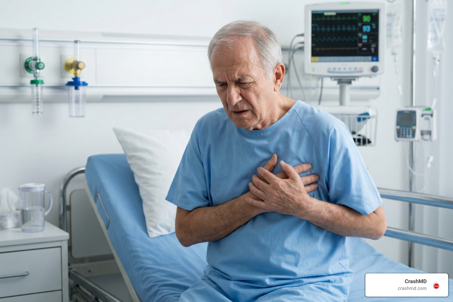
When pain makes breathing difficult, patients involuntarily take shallow breaths—a defensive mechanism known as inspiratory splinting. Learning more about this in our Step-by-Step Guide to Internal Chest Bruising shows how guarding the chest wall can restrict healthy lung function.
Critical Risks of Untreated Cartilage Injury Rib
Failing to address a severe rib cartilage disruption isn’t just uncomfortable—it can become dangerous. When chest wall pain leads to continuous hypoventilation (shallow breathing), the tiny air sacs in the lungs collapse (atelectasis). Stagnant secretions create an ideal environment for secondary bacterial pneumonia, a complication carrying up to a 20% mortality rate in elderly trauma patients.
Furthermore, multi-rib cartilage disruptions can destabilize the thoracic cage, contributing to flail chest mechanics or long-term chronic non-union. As detailed in the Merck Manual rib fracture guide, establishing immediate, effective pain control is essential to maintain proper lung mechanics and avoid respiratory decline.
Treatment Options and Recovery Timeline
Because cartilage receives minimal direct blood supply, healing takes time. A simple cartilage strain or un-displaced tear generally requires 6 to 12 weeks to heal, while severe, displaced costochondral separations can take up to 6 months to completely stabilize.
Conservative management forms the foundation of recovery for most patients:
- Targeted Analgesia: Utilizing non-steroidal anti-inflammatory drugs (NSAIDs) like ibuprofen or acetaminophen to reduce chest wall swelling while preserving respiratory drive.
- Local Nerve Blocks: Administering intercostal nerve blocks or trigger point injections for severe pain that prevents effective deep breathing.
- Avoiding Chest Taping: Rigidly wrapping or taping the chest is strongly discouraged because it constricts chest expansion, worsening atelectasis risks.
- Immune & Respiratory Protection: Preventing respiratory illnesses during early healing is vital, as a single violent cough can re-tear healing cartilage.
For more detailed timelines, review The Ultimate Guide to Rib Cartilage Injury Recovery Timeline.
Surgical Stabilization vs Conservative Care
While non-surgical care is standard, displaced or multi-level costal cartilage fractures can cause persistent pain and reduced breathing capacity. A breakthrough surgical fixation study demonstrated that internal stabilization using low-profile titanium plates significantly improves patient outcomes.
By anchoring custom titanium plates medially to the solid bone of the sternum and laterally to the bony rib shaft (spanning the injured cartilage), surgeons restore structural rigidity. This intervention significantly reduces pain scores, restores chest wall mechanics, and improves forced vital capacity (FVC).
Effective Rehabilitation and Return-to-Play Protocols
Rehabilitation focuses on keeping the lungs clear while gently restoring chest wall mobility and postural strength.
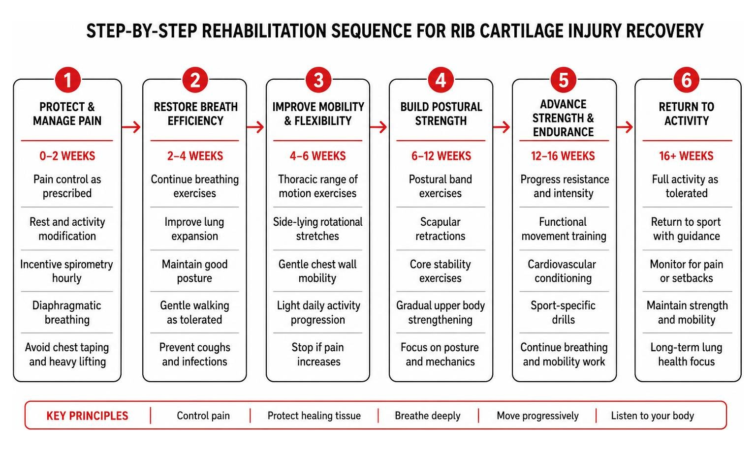
Physical therapy plays a critical role throughout this process. As outlined in the guide Can PT help with rib cartilage injuries?, targeted rehab routines help patients regain functional strength safely:
- Incentive Spirometry & Diaphragmatic Breathing: Patients perform hourly deep breathing exercises, aiming to expand the lower abdomen rather than lifting the painful chest wall.
- Thoracic Range of Motion: Gentle side-lying rotational stretches keep the thoracic spine mobile without loading the healing anterior cartilage.
- Postural Band Exercises: Scapular retractions using light resistance bands strengthen the upper back, reducing stress on the front chest wall.
For competitive athletes in high-impact sports, return-to-play protocols require complete resolution of point tenderness and pain-free chest wall rotation. Athletes returning to contact play should utilize custom-molded chest padding to protect vulnerable costal joints from re-injury.
Frequently Asked Questions About Rib Cartilage Injuries
How long does it take for a rib cartilage injury to heal?
Initial functional recovery typically takes 6 to 12 weeks. However, complete tissue remodeling can take up to 6 months because cartilage is avascular and receives minimal direct blood flow.
Why does a rib cartilage tear not show up on a standard chest X-ray?
Cartilage tissue is radiolucent, meaning X-rays pass through it without creating an image on the film. High-resolution ultrasound, MRI, or 3D reformatted CT scans are necessary to detect non-calcified cartilage tears.
Can a forceful cough or sneeze tear rib cartilage?
Yes. A violent sneeze or severe cough creates high internal pressure and torque across the costal arch, which can tear or subluxate vulnerable cartilage joints—especially if they are already inflamed.
Conclusion
A cartilage injury rib tear is a complex, painful condition that requires expert care. At CrashMD, we guide accident victims across Arizona, Nevada, Texas, Oklahoma, and California through every step of post-collision recovery. We provide specialized pain management, comprehensive medical evaluations, and detailed injury documentation to support both your health and any legal claims resulting from your accident.
If you are suffering from persistent chest pain after a collision, don’t let a normal X-ray leave you without answers. Contact CrashMD for post-accident rib injury evaluation and let our expert medical team help you recover safely.