

When Every Laugh Hurts: Signs of an Injury to Rib Cage Cartilage
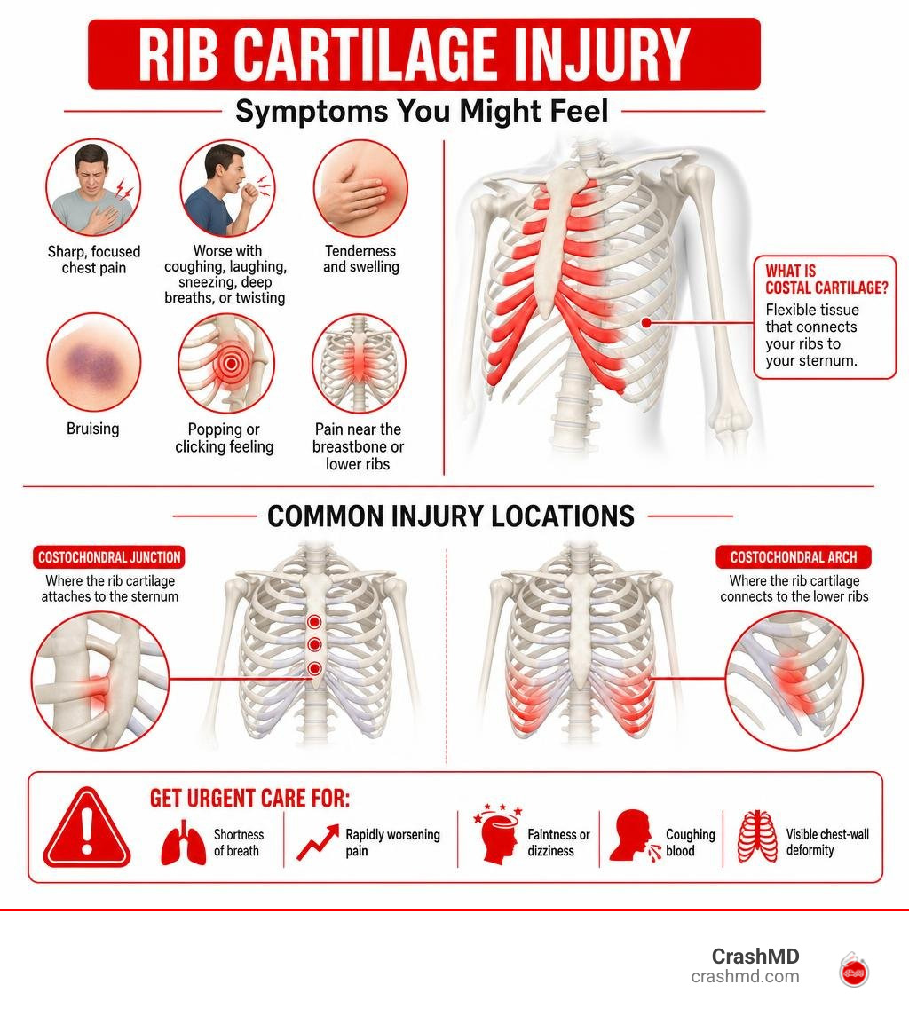
An injury to rib cage cartilage can cause sharp, focused chest pain that gets worse when you laugh, cough, sneeze, take a deep breath, or twist your body. You may also notice tenderness, swelling, bruising, or a popping or clicking feeling near the breastbone or lower ribs. After a car accident, these symptoms deserve medical attention – especially if breathing is painful or difficult.
Costal cartilage is the flexible tissue that helps connect the ribs to the sternum. It can be bruised, torn, separated, or fractured during seatbelt compression, steering-wheel impact, falls, sports collisions, or even forceful coughing. Unlike a broken rib, cartilage damage often does not show on a standard X-ray, so a normal X-ray does not always rule it out.
Seek urgent care for shortness of breath, rapidly worsening pain, faintness, coughing blood, or a visible chest-wall deformity. These can signal a more serious chest injury.
I’m Dr. Adam Savage, and this guide explains what rib cartilage injuries feel like, why they are often missed, and what recovery may involve. Whether you are dealing with an acute cartilage injury to the rib, evaluating how long to recover from rib cartilage injury, or learning how to heal rib cartilage injury, understanding your condition is the first step toward lasting relief.
Anatomy and Common Causes of an Injury to Rib Cage Cartilage
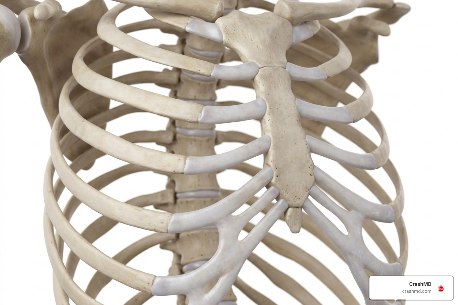
The human rib cage is an engineering marvel. It needs to be rigid enough to shield vital organs like the heart and lungs, yet flexible enough to expand and contract over 20,000 times a day as we breathe. This essential elasticity comes from the costal cartilage—segments of durable hyaline cartilage that connect the bony ribs to the sternum (breastbone).
When blunt chest trauma occurs, the rib cage compresses. If the applied energy exceeds the natural flexibility of these chondral segments, an acute cartilage strain, tear, or fracture takes place.
Understanding Costal Cartilage and Joint Types
The human thoracic cage features 12 pairs of ribs categorized into three distinct groups based on how they connect anteriorly:
- True Ribs (Ribs 1–7): Attach directly to the sternum through individual strips of costal cartilage.
- False Ribs (Ribs 8–10): Do not connect directly to the breastbone; instead, their cartilages merge into a common cartilaginous border known as the costal arch.
- Floating Ribs (Ribs 11–12): Have no anterior cartilaginous connection to the sternum and terminate in the abdominal wall musculature.
These structures form several critical articulations:
- Costochondral Junctions: The points where the bony ribs meet the costal cartilage.
- Chondrosternal (Costosternal) Joints: The junctions where the cartilage attaches directly to the lateral borders of the sternum.
- Manubriosternal and Xiphisternal Junctions: Fibrocartilaginous joints at the top and bottom of the sternum that absorb significant rotational torque.
- Interchondral Articulations: Small synovial connections between the costal cartilages of ribs 6 through 9.
Mechanisms Behind an Acute Injury to Rib Cage Cartilage
Cartilage injuries occur when sudden mechanical forces overcome tissue elasticity. Costal cartilage fractures are estimated to occur in 20% to 42% of patients who sustain high-energy blunt chest trauma (Meteb et al., 2022).
Common mechanisms include:
- Motor Vehicle Collisions: Rapid deceleration causes seatbelts to compress the anterior chest wall violently. Drivers may also experience direct impact against the steering wheel.
- High-Impact Sports: Direct tackles, body checks, or blunt hits in football, rugby, and martial arts compress the costochondral junctions.
- Violent Coughing Fits: Severe bouts of coughing (such as from bronchitis or respiratory infections) can generate enough shearing force from the intercostal muscles to tear cartilage.
- Rotational Torques: Forceful twisting during heavy lifting, golf swings, or rowing can overload the lower costal margin.
A prospective follow-up study on costal cartilage fractures revealed that the most frequent sites of chondral fractures are the 7th rib (21%), the 1st rib (17%), and the 6th rib (15%).
Recognizing the Classic Symptoms and Warning Signs
An injury involving the costal cartilage presents with sharp, localized anterior chest pain. Because cartilage connects directly to the mechanics of breathing, every minor movement of the chest wall can trigger acute discomfort.
Common symptoms include:
- Severe anterior chest pain aggravated by deep inhalation, coughing, sneezing, or laughing.
- Localized tenderness when pressing directly on the ribs near the breastbone.
- Discomfort exacerbated by twisting the torso or bending sideways.
- An audible or palpable “popping,” “clicking,” or snapping sensation during movement.
- Superficial swelling, localized heat, or visible bruising over the sternocostal margin.
Telltale Signs of an Injury to Rib Cage Cartilage vs Rib Fracture
Because ribs and their cartilage sit side-by-side, distinguishing between a bone fracture and a cartilage tear requires careful clinical evaluation.
| Feature | Rib Fracture (Bone) | Rib Cartilage Injury (Chondral) |
|---|---|---|
| Primary Location | Lateral or posterior chest wall | Anterior chest, near the sternum or costal arch |
| X-Ray Visibility | Visible in approximately 50% of cases | Radiolucent (invisible unless calcified) |
| Palpation Sensation | Sharp focal tenderness on bone; bony step-off | Localized tender nodule, swelling, or clicking |
| Mechanical Sensation | Bony crepitus (grating) | Snapping, clicking, or dynamic subluxation |
| Average Healing Time | 6 to 12 weeks | 6 to 12 weeks (up to 6 months for severe tears) |
Red Flag Symptoms and Dangerous Complications
While many cartilage injuries resolve with non-operative care, severe trauma can lead to serious cardiopulmonary complications. Reviewing guidelines on rib trauma evaluation highlights the importance of ruling out internal injuries (Kuo & Kim, 2023).
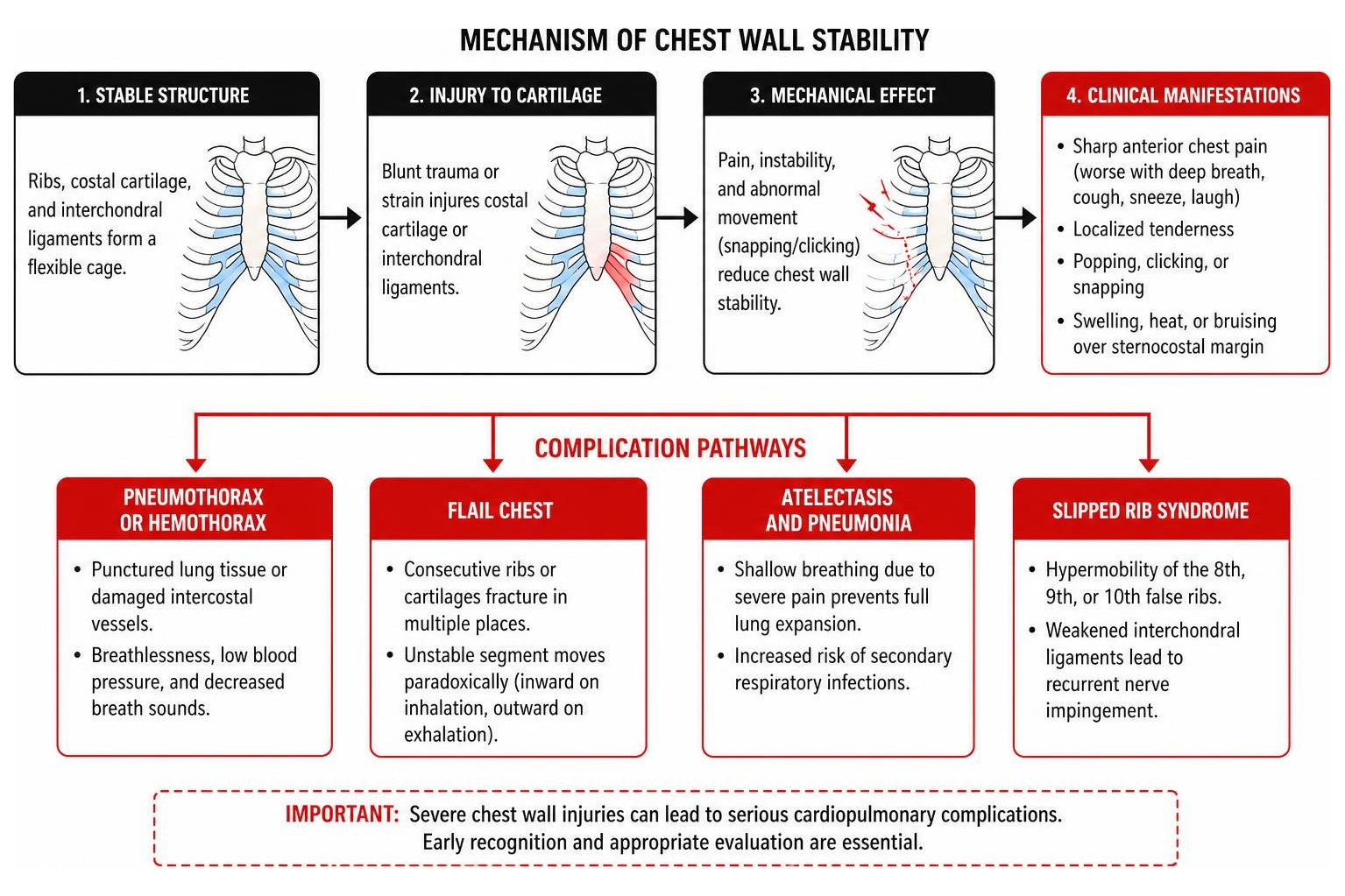
Watch for these warning signs:
- Pneumothorax or Hemothorax: Punctured lung tissue or damaged intercostal vessels causing breathlessness, low blood pressure, and decreased breath sounds.
- Flail Chest: Occurs when consecutive ribs or cartilages fracture in multiple places, creating an unstable floating segment that moves paradoxically (inward on inhalation, outward on exhalation).
- Atelectasis and Pneumonia: Shallow breathing due to severe pain prevents full lung expansion, predisposing patients to secondary respiratory infections.
- Slipped Rib Syndrome: Hypermobility of the 8th, 9th, or 10th false ribs caused by weakened interchondral ligaments, leading to recurrent nerve impingement.
- Chronic Costochondritis or Nonunion: Persistent inflammation or failure of the cartilage ends to bridge stably over time.
Diagnostic Challenges: Why Standard X-Rays Miss Cartilage Tears
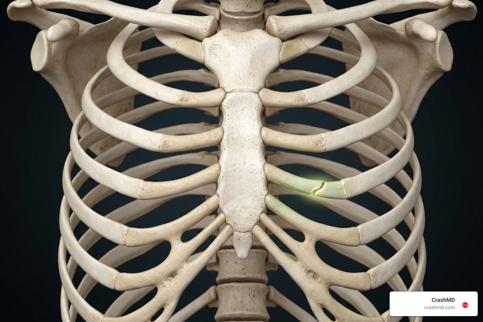
One of the most frustrating aspects of a rib cartilage injury is receiving a “clean” chest X-ray while experiencing severe pain. Plain radiographs rely on calcium density to produce images. Because costal cartilage is composed of uncalcified hyaline matrix, it is radiolucent.
According to comprehensive radiological references on costal cartilage injuries, conventional radiographs cannot visualize cartilage fractures unless significant post-traumatic or age-related calcification is present. In fact, studies show that initial CT evaluations in trauma settings detect costal cartilage fractures only 39.5% of the time unless the radiologist specifically looks for chondral disruptions (Meteb et al., 2022).
High-Resolution Imaging Modalities
When clinical suspicion is high, advanced imaging is necessary:
- Multidetector Computed Tomography (MDCT): Using thin-slice soft-tissue algorithms and 3D volume rendering, CT can clearly show mid-chondral dislocations, sternocostal step-offs, and concurrent internal injuries.
- High-Resolution Dynamic Ultrasound: An exceptional, radiation-free tool for evaluating the anterior chest wall. Dynamic ultrasound can assess the costochondral junction in real time, detecting subtle sliding, tears, and ligamentous instability when the patient deep-breathes or twists.
- Magnetic Resonance Imaging (MRI): The gold standard for soft-tissue contrast. Fat-suppressed T2-weighted and STIR sequences reveal chondral microfractures, joint separations, and bone marrow or cartilage edema that other modalities miss.
Treatment, Rehabilitation, and Expected Healing Timelines
Because cartilage has a limited direct blood supply (relying largely on diffusion from the surrounding perichondrium), healing requires patience and structured care.
Conservative management serves as the foundation:
- Targeted Analgesia: Over-the-counter or prescription NSAIDs help manage both pain and localized chondral inflammation.
- Ice Applications: Applying cold packs for 15–20 minutes at a time during the first 48 to 72 hours minimizes soft-tissue edema.
- Respiratory Care: Using an incentive spirometer and performing gentle hourly deep-breathing exercises prevents atelectasis and pneumonia.
- Avoiding Tight Chest Wraps: While rib binders were once common, modern medical consensus strongly advises against tight chest strapping because it limits lung expansion and dramatically increases pneumonia risk.
If you sustained your injury during a motor vehicle collision, having specialized clinicians evaluate your rib cartilage injury after a car accident ensures your pain is controlled and your injuries are properly documented for your recovery records.
Interventional Options and Surgical Indications
For severe or persistent injuries, additional medical interventions may be necessary:
- Intercostal Nerve Blocks & Trigger Point Injections: Targeted local anesthetic and corticosteroid injections around the affected intercostal nerve can break the cycle of severe pleuritic pain, enabling effective deep breathing.
- Surgical Stabilization and Fixation: Indicated for severe chondral dislocations, flail chest segments, non-healing fractures (nonunion) with chronic pain, or dynamic chest wall deformities. Thoracic surgeons utilize low-profile titanium plates and specialized sutures to bridge and realign disrupted cartilage segments (Kuo & Kim, 2023).
Recovery Phases and Cartilage Repair Timeline
- Weeks 1 to 3 (Acute Phase): Focus is on pain control, gentle breathing exercises, and avoiding all heavy lifting, pushing, pulling, or torso-twisting motions.
- Weeks 4 to 8 (Subacute Phase): Discomfort gradually subsides. Patients begin gentle posture exercises, core stabilization, and light cardiovascular walking.
- Weeks 8 to 12+ (Remodeling Phase): Most isolated cartilage injuries achieve clinical stability as post-traumatic calcifications bridge the gap.
- Up to 6 Months (Complex Injuries): Severe mid-substance tears or multi-level cartilage dislocations may require up to six months for complete mechanical stabilization. Athletes returning to contact sports often benefit from protective chest padding during early competition.
Frequently Asked Questions About Rib Cartilage Damage
How long does it take for injured rib cage cartilage to heal?
Most mild-to-moderate costal cartilage injuries take between 6 and 12 weeks to heal. Because cartilage is avascular, severe tears, separations, or displaced fractures can take up to 6 months for full remodeling and symptom resolution.
Why does an injured rib cartilage hurt more than a broken bone?
Unlike long bones, which can be casted and immobilized, your rib cage moves with every breath, cough, and heartbeat. The continuous motion, combined with the dense network of intercostal nerves running just beneath each rib margin, makes cartilage inflammation feel particularly sharp and persistent.
Can damaged rib cartilage heal properly without surgery?
Yes. The vast majority of rib cartilage injuries heal successfully with conservative management, including pain control, activity modification, and respiratory exercises. Surgery is typically reserved for severe displacement, flail chest, or painful nonunion that fails to improve after several months of conservative treatment.
Conclusion
A rib cartilage injury can turn everyday movements like laughing, coughing, or taking a deep breath into painful challenges. Because costal cartilage is radiolucent, standard X-rays frequently miss these tears, leaving many patients without a clear explanation for their discomfort. Recognizing the symptoms early, securing advanced imaging like high-resolution CT or ultrasound, and following a structured rehabilitation plan are critical steps toward a full recovery.
At CrashMD, we guide accident victims through every stage of healing across Arizona, Nevada, Texas, Oklahoma, Southern California, and Northern California. We specialize in expert post-accident pain management, personalized physical rehabilitation, and thorough medical documentation to protect your health and support your legal case before any settlement decisions. If you are struggling with persistent chest wall pain, schedule an evaluation with our team today to get the answers and relief you deserve.
Scientific References
Meteb M, Abou Shaar B, El-Karim GA, Almalki Y. “Costal cartilage fracture: A commonly missed thoracic injury in trauma patients..” Radiology case reports, 2022. PMCID PMC8571527. Kevin Kuo, Amie M. Kim. “Rib Fracture.” StatPearls Publishing, 2023. NBK541020.