

Why Perforated Bowel Treatment Cannot Wait
Perforated bowel treatment is an emergency. A hole in the small intestine or colon can let bacteria, stool, and digestive fluid leak into the abdomen. Call 911 or go to the emergency room immediately for sudden severe abdominal pain, fever, vomiting, a swollen or rigid belly, fainting, or confusion – especially after a car accident or abdominal injury.
Treatment usually starts with:
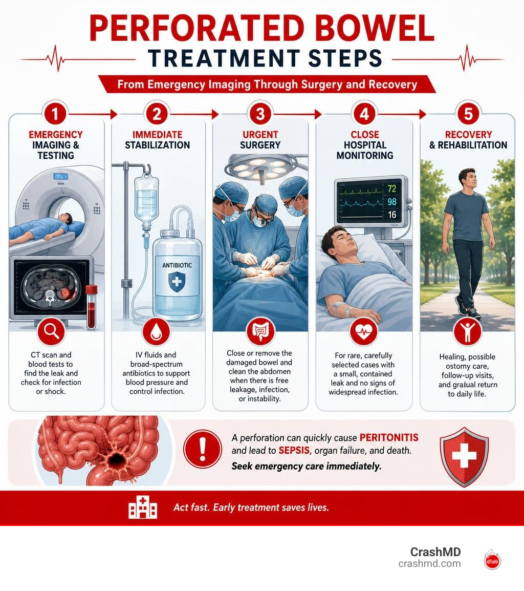
- Emergency CT imaging and blood tests to find the leak and check for infection or shock.
- IV fluids and broad-spectrum antibiotics to support blood pressure and control dangerous infection.
- Urgent surgery to close or remove the damaged bowel and clean the abdomen when there is free leakage, peritonitis, sepsis, or instability.
- Close hospital monitoring for the rare, carefully selected person with a small, contained leak and no signs of widespread infection.
A perforation can quickly cause peritonitis – infection and inflammation of the abdominal lining – and then sepsis, which can lead to organ failure. Most people need prompt surgical care, though certain contained perforations may be managed in the hospital with bowel rest, IV antibiotics, repeat exams, and sometimes drainage of an abscess.
I’m Dr. Adam Savage, and this guide explains what happens next, from emergency diagnosis and surgery to ostomy care, recovery, and accident-related treatment decisions.
Perforated bowel treatment word roundup:
Understanding Bowel Perforation: Causes, Symptoms, and Diagnosis
Common Causes and Accident-Related Trauma
A gastrointestinal perforation occurs when there is a full-thickness breach across the stomach, small intestine, or colon. In emergency rooms and trauma units across Arizona, Nevada, Texas, Oklahoma, and California, blunt abdominal trauma from motor vehicle collisions is a leading mechanical cause. Rapid deceleration can compress the intestines against the spinal column or create a sudden spike in intraluminal pressure, resulting in a blowout injury.
Physical signs like a “seat-belt sign”—bruising across the lower abdomen—warrant urgent clinical attention. We frequently see patients who initially brush off trauma, making our step-by-step guide to assessing abdominal pain after an accident an essential resource. Penetrating injuries, such as stab or gunshot wounds, directly transect intestinal walls.
Beyond physical trauma, underlying medical conditions cause a large percentage of perforations:
- Acute Diverticulitis: Approximately 1% to 2% of diverticulitis patients experience free perforation, while up to 30% develop contained micro-perforations (Jones et al., 2025).
- Peptic Ulcer Disease: Perforations occur in 2% to 10% of cases, primarily involving the duodenum (60%) or stomach (40%).
- Inflammatory Bowel Disease (IBD): Toxic megacolon or deep transmural ulceration leads to perforation in up to 20% of severe ulcerative colitis flares and 1% to 3% of Crohn’s disease cases.
- Iatrogenic Complications: Endoscopic procedures carry a small risk; colonoscopy-related perforation happens in 0.9% to 2% of routine interventions.
- Neoplasms & Foreign Bodies: Colorectal cancer is associated with perforation in 3% to 10% of cases, whereas foreign body ingestion accounts for 1% to 5.6%.
A broader clinical breakdown is detailed in our detailed guide to causes of perforated bowel urgency as well as the acute gastrointestinal perforation overview.
Clinical Symptoms and Emergency Warning Signs
When intestinal contents spill into the sterile peritoneal cavity, chemical irritation rapidly gives way to bacterial infection. The hallmark symptom is sudden, excruciating abdominal pain that often worsens with movement or light palpation.
Key physical warning signs include:
- Involuntary Guarding and Rigidity: The abdominal wall muscles reflexively tense into a board-like firmness to shield inflamed internal organs.
- Rebound Tenderness: Sharp, intense pain experienced upon the sudden release of manual pressure on the abdomen.
- Systemic Sepsis Indicators: Tachycardia (heart rate > 90 bpm), tachypnea (rapid breathing), high-grade fever, hypothermia, or profound hypotension (low blood pressure).
- Gastrointestinal Distress: Persistent nausea, bilious vomiting, progressive abdominal distension, and complete cessation of bowel sounds or flatulence.
Recognizing these manifestations prevents dangerous delays. For an exhaustive breakdown, consult our 7 devastating symptoms of perforated bowel and the ultimate checklist for perforated bowel emergency signs. The systemic threat is further outlined in the Sepsis Alliance guide to bowel perforation complications.
Diagnostic Testing and Emergency Imaging

Accurate and swift diagnostic workup is critical for determining whether non-operative management or surgical intervention is required.
Initial triage involves an upright chest radiograph or a left lateral decubitus abdominal X-ray. These plain films identify free subdiaphragmatic air (pneumoperitoneum) in 50% to 70% of perforation cases. However, the absence of free air on plain X-rays does not rule out a perforation.
The gold standard confirmatory test is an abdominal and pelvic CT scan with intravenous (IV) contrast. CT imaging provides exceptional diagnostic sensitivity by detecting subtle extraluminal gas, localized fluid collections, bowel wall thickening, and mesenteric fat stranding, precisely pinpointing the perforation site. Oral contrast is used cautiously or substituted with water-soluble agents to prevent barium-induced chemical peritonitis.
Laboratory diagnostics complement imaging:
- Complete Blood Count (CBC): Reveals pronounced leukocytosis (elevated white blood cell count with left shift) or leukopenia in severe sepsis.
- Serum Lactate: Serves as a vital biomarker for tissue hypoperfusion, anaerobic metabolism, and occult septic shock.
- Metabolic & Coagulation Panels: Evaluates renal function, electrolyte derangements, and coagulation profiles prior to emergency surgery.
For a deep dive into diagnostic protocols, review the AMBOSS diagnostic guidelines on gastrointestinal perforation.
Comprehensive Perforated Bowel Treatment Pathways
Non-Surgical Perforated Bowel Treatment and Conservative Care
Not every gastrointestinal perforation requires an immediate trip to the operating room. In strictly selected clinical scenarios, conservative, non-operative management is both safe and effective (Levack et al., 2014).
Non-surgical care is considered exclusively when all of the following criteria are met:
- The perforation is small and anatomically contained (e.g., walled off by adjacent omentum or mesentery).
- The patient is hemodynamically stable without evidence of severe sepsis or septic shock.
- Diffuse peritonitis is entirely absent during serial physical examinations.
Conservative management involves strict bowel rest (NPO status), aggressive IV crystalloid hydration, and broad-spectrum intravenous antimicrobial therapy (Levack et al., 2014). If a localized intra-abdominal abscess develops (typically $ge 3text{ cm}$), interventional radiologists can perform CT-guided percutaneous drain placement to evacuate infected fluid without surgical incision (Jones et al., 2025). Patients require continuous monitoring in an acute care setting with serial blood work and repeat CT scans; any signs of clinical deterioration trigger immediate conversion to surgery (Jones et al., 2025). See the StatPearls review on bowel perforation clinical management for deeper insights.
Surgical Approaches in Perforated Bowel Treatment

When free perforation, generalized peritonitis, or clinical instability is present, emergent surgical exploration is required. The surgical strategy depends on the underlying etiology, anatomical location, degree of peritoneal contamination, and the patient’s physiological status.
- Primary Suture Repair: For clean, localized traumatic tears or small perforated peptic ulcers, simple closure using interrupted sutures (often reinforced with an omental pedicle, known as a Graham patch) is preferred.
- Segmental Bowel Resection: When extensive necrosis, large diverticular phlegmon, severe ischemia, or underlying malignancy is present, the compromised intestinal segment is resected.
- Laparoscopic vs. Open Laparotomy: While emergent exploratory laparotomy through a midline incision remains the gold standard for diffuse fecal peritonitis, diagnostic laparoscopy and minimally invasive repair are increasingly utilized in stable patients with localized contamination (Nascimbeni et al., 2021).
- Peritoneal Lavage & Debridement: Copious warm saline irrigation is performed to evacuate all purulent exudate, enteric debris, and fibrinous peel, significantly reducing the intra-abdominal bacterial load.
Detailed surgical recommendations can be found in the multidisciplinary guidelines on perforated diverticulitis management.
Damage Control Surgery and Critical Resuscitation
In critically ill patients presenting with septic shock, hypothermia, acidosis, and coagulopathy (the “lethal triad”), definitive prolonged reconstructive surgery can be fatal. In these high-risk scenarios, surgeons utilize Damage Control Surgery (DCS) (Harikrishnan et al., 2025).
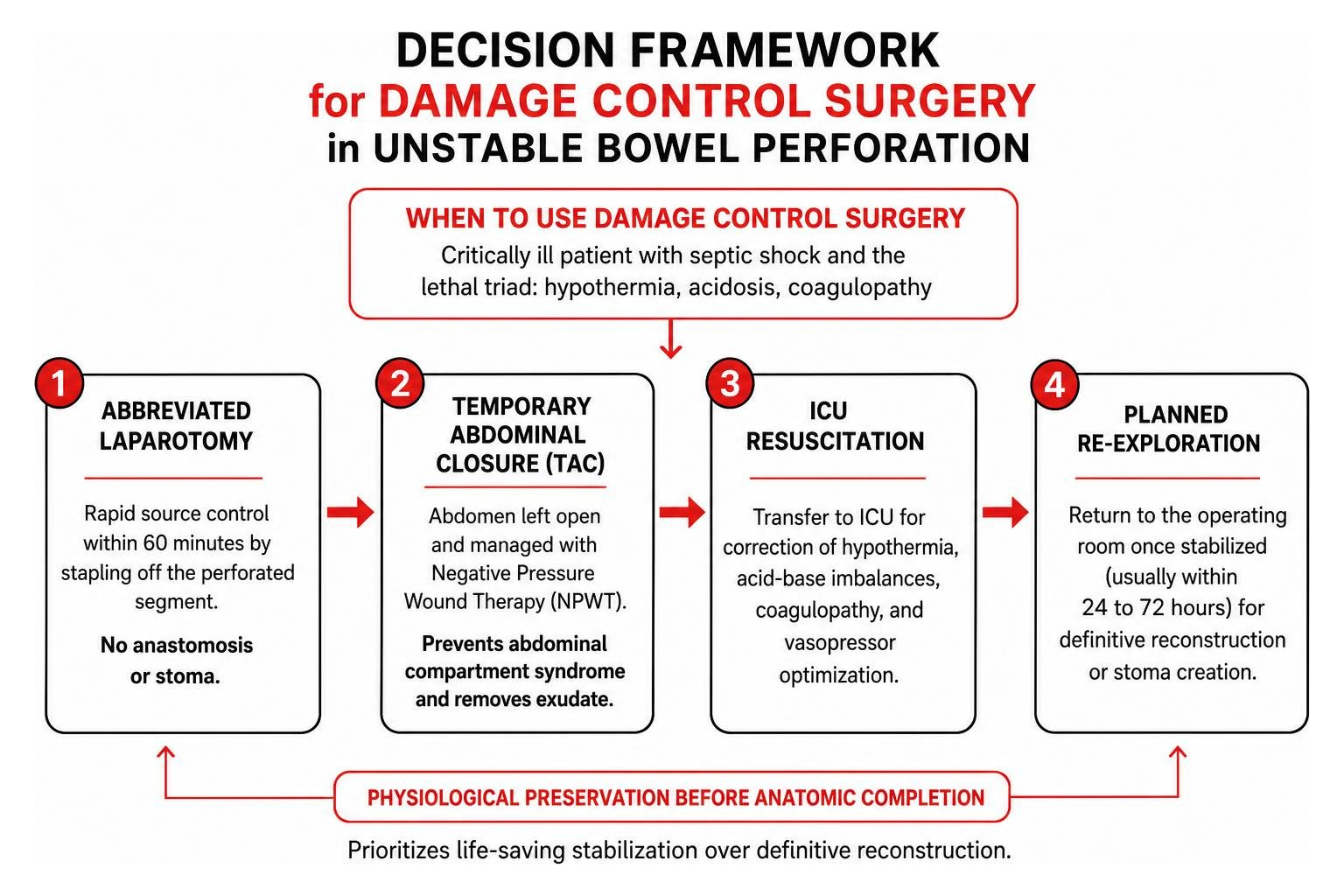
DCS prioritizes physiological preservation over anatomic completion:
- Abbreviated Laparotomy: Rapid surgical source control is achieved within 60 minutes by stapling off the perforated segment without performing an anastomosis or stoma.
- Temporary Abdominal Closure (TAC): The abdomen is left open and managed using Negative Pressure Wound Therapy (NPWT) to prevent abdominal compartment syndrome and remove ongoing exudate (Harikrishnan et al., 2025).
- ICU Resuscitation: The patient is transferred to the intensive care unit for correction of hypothermia, acid-base imbalances, coagulopathy, and vasopressor optimization.
- Planned Re-exploration: Once stabilized (usually within 24 to 72 hours), the patient returns to the operating room for definitive reconstruction or stoma creation (Harikrishnan et al., 2025).
Clinical evidence demonstrating improved survival is outlined in the systematic review on damage control surgery.
Ostomy Surgery, Antibiotic Therapy, and Critical Interventions
Colostomy and Ileostomy: Indications and Stoma Management
When inflamed or contaminated bowel tissues are too fragile to safely sew back together, performing a primary anastomosis carries a high risk of leaking. In these cases, surgeons divert the stool stream by creating a stoma on the abdominal wall.
- Hartmann’s Procedure: Involves resecting the diseased colonic segment (commonly the sigmoid colon), oversewing the rectal stump, and bringing the proximal colon out as an end colostomy.
- Loop Ileostomy: A loop of the small intestine is brought through the skin to divert fecal contents away from a vulnerable distal repair, allowing downstream tissues to heal.
Stomas can feel overwhelming, but for most patients with trauma or perforated diverticulitis, they are temporary. Reversal surgeries are typically scheduled 3 to 6 months post-discharge after systemic inflammation subsides and bowel integrity is radiographically confirmed. Learn more via the Cleveland Clinic clinical overview on gastrointestinal perforation surgery.
Role of Intravenous Antibiotics and Infection Control
Empiric broad-spectrum intravenous antibiotic therapy must be initiated immediately upon suspicion of bowel perforation. Regimens must cover enteric Gram-negative bacilli (e.g., Escherichia coli, Klebsiella), Gram-positive cocci (Enterococcus), and obligate anaerobes (Bacteroides fragilis).
Common antimicrobial regimens include beta-lactam/beta-lactamase inhibitors (piperacillin-tazobactam), third-generation cephalosporins paired with metronidazole, or carbapenems (meropenem) for septic shock. Once intraoperative microbial cultures and sensitivities return, targeted de-escalation prevents drug resistance.
| Feature | Conservative Medical Management | Urgent / Emergent Surgical Management |
|---|---|---|
| Primary Indication | Contained micro-perforation, stable vital signs, localized phlegmon | Free perforation, diffuse peritonitis, hemodynamic instability |
| Interventions | Strict NPO, IV antibiotics, percutaneous drain ($ge 3text{ cm}$) | Laparoscopy or Laparotomy, primary repair, resection $pm$ stoma |
| ICU Requirement | Rare (unless significant underlying comorbidities) | Common (especially with damage control surgery or septic shock) |
| Dietary Timeline | Gradual oral sips upon clinical and lab improvement | NPO with NG decompression; oral diet after bowel motility returns |
| Monitoring Needs | Serial abdominal exams, daily labs, repeat CT at 7–10 days | Wound assessment, drain monitoring, stoma output tracking |
Recovery Timeline, Potential Complications, and Prognosis
Postoperative Rehabilitation and Healing Milestones
Recovery following emergency bowel surgery is a phased process that begins in the hospital and continues for months at home.
In the immediate postoperative period (Days 1–4), care focuses on nasogastric tube decompression to prevent stomach distension, aggressive fluid management, and multimodal pain control to minimize opioid-induced paralytic ileus. Early ambulation within 24 hours of extubation reduces the risk of deep vein thrombosis (DVT) and stimulates gastrointestinal motility.
Patients gradually transition from clear liquids to a low-residue, high-protein diet as bowel sounds and flatus return. Full physical recovery generally takes 6 to 12 weeks. During this time, patients must adhere to strict lifting restrictions (< 10–15 lbs) to prevent incisional hernias and participate in physical rehabilitation to restore core abdominal strength.
Managing Complications: Sepsis, Abscesses, and Fistulas
Because gastrointestinal perforation involves bacterial spillage, the risk of secondary complications remains elevated throughout recovery:
- Tertiary Peritonitis & Abscesses: Persistent fever and leukocytosis often indicate retained intra-abdominal fluid collections requiring percutaneous drainage.
- Anastomotic Leak: Breakdown of surgical bowel connections causes renewed peritonitis, necessitating emergency re-operation.
- Enterocutaneous Fistula: An abnormal tract forming between the intestinal lumen and the skin surface, requiring complex nutritional and wound management.
- Adhesive Small Bowel Obstruction: Internal scar tissue bands can kink the bowel months or years later.
For individuals recovering from vehicle impacts, specialized rehabilitation and injury tracking are critical. Explore our car accident intestinal perforation recovery guidance and learn about your immediate care after an accident options to safeguard your long-term physical and legal recovery.
Frequently Asked Questions About Perforated Bowel Surgery
Can a perforated bowel heal without surgical intervention?
Yes, but only in strictly selected cases. If the perforation is microscopic, walled off by surrounding abdominal fat (omentum), and the patient displays stable vital signs with zero generalized peritonitis, hospital-based conservative management with IV antibiotics and bowel rest can allow the tissue to seal itself (Levack et al., 2014).
What is the expected recovery time after bowel perforation surgery?
Hospital stays typically range from 5 to 14 days depending on surgical complexity. Full recovery at home requires 6 to 8 weeks of restricted physical activity, specialized dietary modifications, and wound care before resuming strenuous labor or exercise.
How does emergency surgery prevent life-threatening sepsis?
Surgery provides immediate “source control.” By closing the hole, removing necrotic tissue, and washing out leaked stool and bacteria from the abdominal cavity, surgery halts the ongoing bacteremia and suppresses the overwhelming systemic inflammatory cascade that drives multi-organ failure.
Conclusion
A perforated bowel is a critical medical emergency that requires swift diagnosis, decisive intervention, and comprehensive post-surgical care. Whether managed through specialized conservative protocols or emergent operative repair, timely medical action is the single most important factor determining complete recovery.
For accident victims navigating severe abdominal trauma, medical care extends beyond acute surgery. At CrashMD, we advocate for patients across Arizona, Nevada, Texas, Oklahoma, and California by providing expert trauma rehabilitation, comprehensive pain management, and thorough post-accident medical documentation. Navigating complex post-trauma recovery requires an experienced multidisciplinary team—ensuring you regain your physical health, document your injuries accurately, and protect your long-term wellness.
Scientific References & Clinical Citations
Levack MM, Madariaga ML, Kaafarani HM. “Non-operative successful management of a perforated small bowel diverticulum..” World journal of gastroenterology, 2014. PMCID PMC4277989. Mark W. Jones, Sarang Kashyap, Bradley Boget, Christopher P. Zabbo. “Bowel Perforation.” StatPearls Publishing, 2025. NBK537224. Nascimbeni R, Amato A, Cirocchi R, Serventi A, Laghi A, Bellini M, Tellan G, Zago M, Scarpignato C, Binda GA. “Management of perforated diverticulitis with generalized peritonitis. A multidisciplinary review and position paper..” Techniques in coloproctology, 2021. PMCID PMC7884367. Harikrishnan S, Neelakantan Ramaswamy S, Agarwal Y, Bhatt NK, Chand S, Bendagiri Matam M, Mohammed L. “Role of Damage Control Surgery in Perforated Diverticulitis Management: A Systematic Review..” Cureus, 2025. PMCID PMC12417249.