

How Pulmonary Laceration Appears on Trauma Imaging
Pulmonary laceration radiology starts with finding a tear in the lung that forms a round or oval space filled with air, blood, or both. On CT, look for a well-defined cavity, an air-fluid level, or a blood-filled mass within an area of lung bruising (contusion). A chest X-ray can miss the injury early because the surrounding contusion may hide it. In a stable trauma patient, chest CT is the best test to detect and define the laceration.
A pulmonary laceration can happen after a car crash, fall, or other blunt force to the chest. The lung’s elastic tissue pulls back after it tears, which is why these injuries often look like cavities rather than straight lines on imaging. They commonly occur alongside contusions, rib fractures, pneumothorax, or bleeding around the lung.
On follow-up imaging, the cavity may become easier to see as the surrounding bruise clears. Most lacerations gradually shrink and resolve over roughly 3 to 5 weeks, though larger injuries or positive-pressure ventilation can delay healing or enlarge an air-filled cavity.
I’m Dr. Adam Savage, and the sections ahead explain how clinicians read these findings, separate them from other lung injuries, and watch for complications that need urgent care.
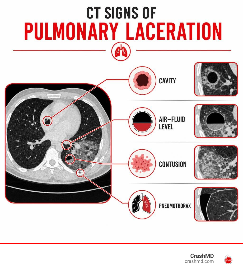
Discover more about pulmonary laceration radiology:
Pulmonary Laceration Radiology: Plain Radiography vs. Multidetector CT
Pulmonary lacerations occur in up to 70% of blunt thoracic trauma cases, making parenchymal disruption a frequent finding in severe motor vehicle collisions. Evaluating these injuries begins with understanding the diagnostic limits of plain radiographs versus high-resolution cross-sectional imaging [[mcdoi3367d42ca7b9:INLINE]]. While reading a pulmonary contusion guide highlights alveolar hemorrhage, lacerations represent actual structural tears within the parenchyma.
Detailed reviews such as Pulmonary contusions and lacerations: CT findings show that multidetector computed tomography (MDCT) achieves near 100% sensitivity for identifying parenchymal trauma. In contrast, plain chest radiography detects only about 37.5% to 50% of these lesions during initial trauma resuscitation.
Chest Radiography Findings and Obscuration Pitfalls
On an initial supine or portable anteroposterior (AP) chest radiograph, pulmonary lacerations are frequently invisible. Around 50% of lacerations go unnoticed on initial plain films because dense parenchymal consolidation from adjacent alveolar hemorrhage completely obscures the underlying tear.
When evaluating severe blunt impact, clinicians managing internal chest bruising and trauma evaluation must maintain suspicion for occult tears. Radiographic clues often rely on indirect signs:
- Delayed Cavitation: As surrounding hemorrhagic consolidation resorbs over 48 to 72 hours, an air-filled lucency or air-fluid level gradually unmasks.
- Deep Sulcus Sign: On supine radiographs, an abnormally deep, radiolucent costophrenic angle indicates an associated occult pneumothorax.
- Pleural Fluid Collections: Rapidly accumulating hemopneumothorax often signals an underlying parenchymal disruption.
Multidetector CT Imaging Features and Diagnostic Superiority
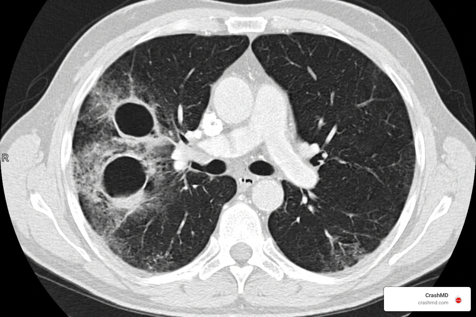
Thin-slice MDCT with intravenous contrast is the gold standard for thoracic trauma assessment. Routine reconstructions in dedicated lung windows (width ~1500 HU, level ~-600 HU) clearly distinguish intact, contused lung from structural tears.
Published cases in the Pulmonary lacerations imaging review demonstrate several classic CT patterns:
- Ovoid or Spherical Cavities: Due to elastic recoil pulling the margins outward, parenchymal tears present as round or oval spaces rather than linear clefts.
- “Swiss Cheese” Pattern: Multiple small, clustered, air-filled cysts surrounded by ground-glass opacities.
- Air-Fluid Levels: Dependent blood filling the bottom of an air-filled space (haemato-pneumatocele).
- Active Vascular Contrast Extravasation: A localized “blush” of high-attenuation contrast within the laceration cavity, signaling active arterial or venous hemorrhage that may warrant urgent transcatheter embolization.
Classification of Traumatic Lung Tears and Morphology
The biomechanical forces involved in blunt chest trauma dictate where and how lung parenchyma tears. When external compression abruptly deforms the thoracic cage, lung tissue stretches past its tensile limits. Surrounding parenchyma snaps back via inherent elastic recoil, transforming the tear into a gas- or blood-filled cavity.
As outlined in the Pulmonary laceration reference article, the Wagner classification framework categorizes these injuries into four distinct types based on the mechanism of trauma and CT morphology [[mcpmcidbc36c49077a8:INLINE]].
| Wagner Type | Injury Mechanism | Typical Anatomic Location | Classical CT Imaging Appearance |
|---|---|---|---|
| Type I | Rapid compressive force / compression rupture | Deep, central lung parenchyma | Round or oval cavity (2–8 cm), often with air-fluid level |
| Type II | Sudden shearing displacement | Paravertebral lower lobe | Tubular or elongated air-fluid collection along the spine |
| Type III | Direct penetrating puncture from rib fragment | Peripheral lung adjacent to fractured rib | Small, peripheral cavity abutted by fractured bone edges |
| Type IV | Shear stress across pre-existing pleural adhesions | Subpleural periphery at site of prior adhesion | Hemispherical or crescentic peripheral cavity; often leads to pneumothorax |
Interpreting Pulmonary Laceration Radiology Patterns Across Wagner Types I-IV
Understanding the specific imaging characteristics of each Wagner subtype aids in injury mapping:
- Type I (Compression Rupture): The most frequent variant. Occurs when high-energy blunt force compresses the chest against a closed glottis, rupturing deep alveolar walls. It typically produces a 2 to 8 cm central cavity.
- Type II (Compression Shear): Results from severe lower-chest compression that forcibly displaces the lower lobe across the rigid thoracic vertebrae. CT shows a distinctive tubular laceration adjacent to the spine.
- Type III (Rib Penetration): A displaced rib fracture directly punctures the peripheral lung surface. CT bone windows confirm jagged cortical edges projecting into the subpleural space.
- Type IV (Pleuropulmonary Adhesion Tears): Sudden deceleration pulls strongly against fixed fibrous adhesions, ripping the lung periphery and frequently causing large, persistent pneumothoraces.
Trauma teams also utilize the American Association for the Surgery of Trauma (AAST) lung injury grading scale to score severity, ranging from minor unilateral contusions (Grade I) to massive parenchymal disruption involving major hilar vessels (Grade V).
Differentiating Lacerations from Contusions, Hematomas, and Pseudocysts
Distinguishing post-traumatic parenchymal injuries on cross-sectional imaging ensures accurate prognosis. Reviewing pulmonary contusion radiology imaging features provides helpful context for differentiating these overlapping entities:
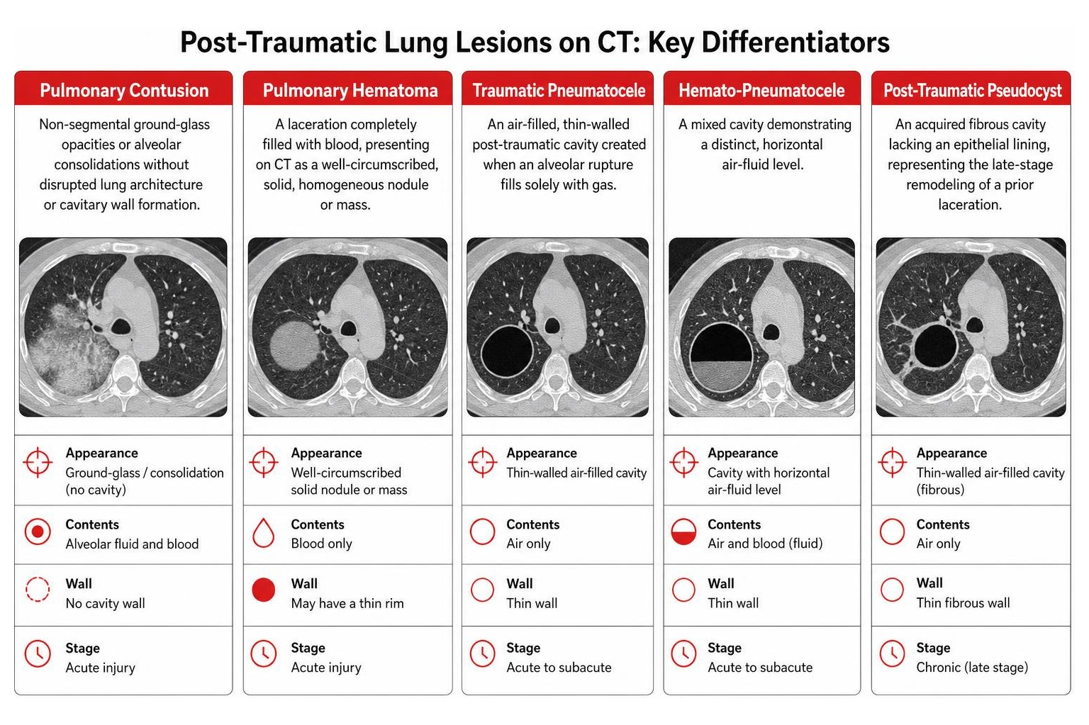
- Pulmonary Contusion: Non-segmental ground-glass opacities or alveolar consolidations without disrupted lung architecture or cavitary wall formation.
- Traumatic Pneumatocele: An air-filled, thin-walled post-traumatic cavity created when an alveolar rupture fills solely with gas.
- Pulmonary Hematoma: A laceration completely filled with blood, presenting on CT as a well-circumscribed, solid, homogeneous nodule or mass.
- Hemato-Pneumatocele: A mixed cavity demonstrating a distinct, horizontal air-fluid level.
- Post-Traumatic Pseudocyst: An acquired fibrous cavity lacking an epithelial lining, representing the late-stage remodeling of a prior laceration.
Age-Related Patterns and Clinical Evolution Over Time
Understanding patient-specific anatomy and monitoring temporal changes helps prevent diagnostic errors. Early assessment also highlights common pulmonary contusion risks and complications that occur alongside parenchymal tears.
Age-Related Variations in Pulmonary Laceration Radiology: Pediatric vs. Adult Findings
Age alters the radiological presentation of thoracic trauma due to differences in chest wall compliance:
- Pediatric Patients: Children possess highly compliant, cartilaginous thoracic cages. Significant kinetic energy transfers directly into the pulmonary parenchyma without fracturing bones. Consequently, pediatric lacerations frequently occur without overlying rib fractures and often feature subpleural sparing.
- Adult Patients: Adult thoracic cages are rigid and more calcified. Lacerations in adults strongly correlate with multiple displaced rib fractures, flail chest segments, larger cavity dimensions (averaging ~2.8 cm compared to ~1.9 cm in pediatric cohorts), and higher rates of concomitant hemothorax.
Because children are more radiosensitive, pediatric imaging strategies prioritize optimized low-dose MDCT protocols.
Serial Imaging Progression and Positive Pressure Ventilation Effects
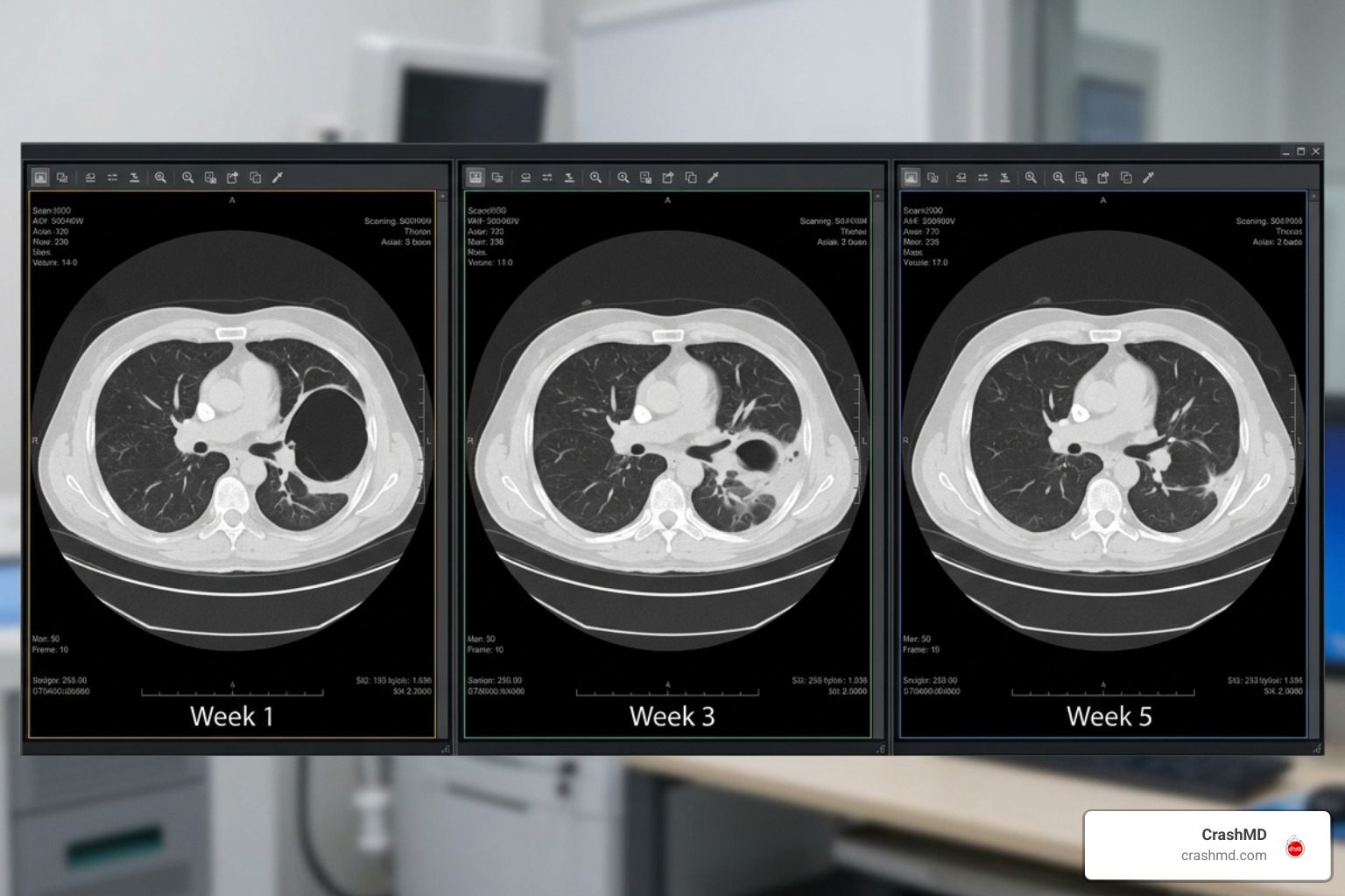
Uncomplicated pulmonary lacerations generally follow a predictable healing timeline. The surrounding contusion peaks at 48 to 72 hours and resolves within 7 to 14 days, while the underlying laceration shrinks and heals over 3 to 5 weeks, ultimately leaving a small fibrotic scar.
However, mechanical ventilation can alter this process:
- Positive Pressure Ventilation (PPV): Mechanical ventilation delivers continuous intrathoracic pressure. This force can push air through torn bronchioles into the laceration, creating a “ball-valve” mechanism.
- Expanding Pneumatoceles: A stable 2 cm cavity can rapidly inflate into a giant, tension pneumatocele.
- Tension Pneumothorax: Progressive expansion risks bursting the cavity into the pleural space, precipitating life-threatening tension physiology. Ventilator parameters must be carefully managed with low tidal volumes and titrated positive end-expiratory pressure (PEEP) to avoid barotrauma.
Complications, Differential Diagnoses, and Management Protocols
Most patients with isolated pulmonary lacerations recover with conservative supportive care, including oxygen therapy, pain management, and pulmonary hygiene. However, severe trauma requires vigilant monitoring.
According to trauma literature in Imaging manifestations of thoracic injury, roughly one-third of thoracic injury patients who require surgical intervention undergo lung resection. Resection carries a stepwise escalation in mortality:
- Wedge Resection: ~19% mortality
- Lobectomy: ~27% mortality
- Pneumonectomy: ~53% mortality
Given these surgical risks, non-operative management guided by serial imaging remains the preferred pathway whenever clinically feasible.
Identifying High-Risk Complications: Bronchopleural Fistula and Active Bleeding
Radiologists and trauma surgeons look for specific imaging signs to identify complications requiring urgent intervention:
- Bronchopleural Fistula (BPF): A structural communication between a torn bronchus and the pleural space. CT demonstrates direct continuity between an airway, the laceration, and a persistent pneumothorax accompanied by continuous bubbling in the chest drain.
- Active Parenchymal Hemorrhage: Contrast-enhanced CT reveals active extravasation (a pooling contrast jet measuring 85–350+ HU). While stable tears resolve spontaneously, expanding hematomas with active extravasation may require prompt transcatheter arterial embolization (TAE).
- Secondary Pulmonary Abscess: Superinfection of a fluid-filled hematoma, appearing on CT as a thick, irregular, contrast-enhancing wall with new internal gas bubbles and surrounding consolidation, accompanied by clinical fever and leukocytosis.
Differential Diagnosis of Cavitary Lesions in the Trauma Bay
When a cavitary lung lesion appears on an acute chest CT, clinicians must distinguish traumatic tears from other medical conditions. Documenting these differences is also essential when assigning the appropriate ICD-10 coding for pulmonary contusion and laceration.
- Post-Contusion Pneumonia Cavitation: Appears 5 to 10 days post-injury due to necrotizing secondary bacterial infection, unlike immediate traumatic lacerations seen on day one.
- Pre-Existing Bullae or Emphysematous Blebs: Extremely thin walls (<1 mm) lacking surrounding ground-glass hemorrhage or contusion, often located at the lung apices.
- Cavitary Tuberculosis or Fungal Disease: Typically features chronic apical fibronodular changes, upper lobe predominance, and tree-in-bud opacities without an acute trauma history.
- Septic Pulmonary Emboli: Multiple, wedge-shaped peripheral nodules exhibiting variable stages of central cavitation, frequently associated with infective endocarditis or infected central lines.
Frequently Asked Questions About Pulmonary Lacerations
How long does it take for a pulmonary laceration to heal on follow-up imaging?
Pulmonary lacerations typically resolve over 3 to 5 weeks. Serial CT surveillance demonstrates gradual shrinkage of the air-fluid collection, leaving a small linear or stellate fibrotic scar. Healing can take several months in patients with severe ARDS, secondary infection, or prolonged mechanical ventilation.
Why are pulmonary lacerations often missed on initial chest X-rays?
Approximately 50% of lacerations are obscured on admission chest radiographs because dense alveolar consolidation from concurrent pulmonary contusion surrounds and masks the parenchymal tear. As the contusion resolves over 48 to 72 hours, the underlying gas- or fluid-filled cavity becomes visible.
What imaging signs indicate the need for urgent thoracic surgery after a lung laceration?
Urgent surgical exploration or interventional embolization is indicated when imaging and clinical monitoring demonstrate active contrast extravasation, a rapidly expanding tension pneumatocele causing mediastinal shift, massive unevacuated hemothorax (>1500 mL output), or a large central bronchopleural fistula that fails conservative chest tube drainage.
Conclusion
Accurate interpretation in pulmonary laceration radiology requires recognizing that the lung’s natural elasticity transforms tears into round cavities, hematomas, or hemato-pneumatoceles rather than linear defects. While initial radiographs frequently miss these injuries due to surrounding contusion, high-resolution multidetector CT provides definitive detection, classification, and complication monitoring.
Following acute chest trauma, evaluating injuries and tracking recovery are critical first steps. For comprehensive post-accident care and support understanding car accident pulmonary contusion symptoms and recovery, our team at CrashMD provides specialized pain management, objective diagnostic documentation, and coordinated care across our clinics in Arizona, Nevada, Texas, Oklahoma, Southern California, and Northern California.
Scientific References
Carboni Bisso I, Gemelli NA, Barrios C, Las Heras M. “Pulmonary laceration..” Trauma case reports, 2021. PMCID PMC8010856. Camilo Dallagnol, Juan M. F. Alcala, Rafaelle M. Vargas, Gustavo R. dos Santos, Dante L. Escuissato. “Pulmonary contusions and lacerations: CT findings that every radiologist must know.” Chinese Journal of Academic Radiology, 2024. DOI 10.1007/s42058-023-00133-w.